Tattoo
A tattoo may occur accidentally or deliberately for cosmetic and decorative purposes. Accidental tattooing results from unintentional deposition of exogenous pigmented substances such as asphalt, carbon, or graphite (pencils) within injured skin (Fig. 94.5). Automobile, bicycle, and skating accidents as well as puncture wounds are the most common causes of traumatic tattoos. In decorative tattooing, the pigment is introduced into the dermis via needles or a tattoo gun to create various designs, groupidentity symbols, or writings. Cosmetic tattoos are also done to define the contours of lips and eyebrows, permanently replace eyeliner, make eyebrows appear fuller (via microblading), or camouflage an abnormality in skin color. An iatrogenic tattoo may remain after the use of ferrous subsulfate (Monsel’s solution) for hemostasis. Other hemostatic agents, e.g. aluminum chloride, may cause a foreign body reaction histologically (see below), but do not lead to discoloration of the skin.
Decorative tattoos often contain a number of pigments that are combined to create different hues, but the composition of tattoo inks has not been standardized. The pigments may be inorganic salts of metals, such as mercury in cinnabar (red, becoming historical), cobalt
(blue), chromium (green), cadmium (yellow, red), ferric hydrate (sienna) and manganese (purple), or organic preparations.
Clinical features
Foreign body reactions may occur within a few weeks of the tattoo placement, but have been reported decades later. Most commonly, erythematous papules, nodules, or plaques are seen (Fig. 94.6), but lesions may be verrucous, lichenoid, pseudolymphomatous, or eczematous. The reactions are usually confined to the site of the tattoo, with the degree of tenderness and erythema reflecting the intensity of the associated inflammation. “Allergic” reactions most frequently occur within the red-colored areas of the tattoo (Fig. 94.7), while sarcoidal
PAS, periodic acid Schiff.
This type of tattoo responds favorably to Q-switched laser therapy.
reactions favor sites of black color. In some patients, granulomatous papulonodules within a tattoo can be a manifestation of cutaneous or systemic sarcoidosis. Rarely, a photoallergic reaction, presenting as pruritic, inflamed nodules, occurs in red or yellow tattoos containing cadmium sulfide following exposure to UV light.
Verrucous lesions and pseudoepitheliomatous hyperplasia can also develop within tattoos and may be misdiagnosed as human papillomavirus (HPV) infection or squamous cell carcinoma (SCC). The development of keratoacanthomas or SCCs within tattoos is rare. Tattoo pigments may migrate to the regional lymph nodes and at the time of lymph node sampling may clinically mimic metastatic melanoma.
Development of inflammation within older tattoos is occasionally triggered by a new tattoo that contains the same pigments. In the Rush phenomenon, granulomatous papulonodules initially develop in the new tattoo then within older tattoos, with sarcoidosis representing a risk factor.
Pathology
Different patterns of an inflammatory response can be elicited, including sarcoidal, foreign body granulomatous, lichenoid, eczematous, and pseudolymphomatous. The majority of tattoo pigments appear black in H&E-stained sections, regardless of the color of the pigment (see Fig. 72.23B). Occasionally, colors other than black are seen (see below). The pigment is deposited in the upper and mid dermis, both extracellularly between collagen bundles and around blood vessels as well as a smaller amount within the cytoplasm of macrophages.
The pigment deposits in professional tattoos are relatively small and homogeneous in size and shape, while in traumatic tattoos they are larger and vary in size and shape. Coarse, granular brown pigment within macrophages and between collagen fibers are seen in iatrogenic tattoos due to ferrous subsulfate (Monsel’s solution; Fig. 94.8).
Diagnosis and differential diagnosis
The presence of inflammatory lesions confined to the site of a tattoo suggests the diagnosis. Inoculation of certain infectious agents (e.g. atypical mycobacteria) may mimic pigment-induced inflammation within a tattoo (Table 94.4). In addition to hypertrophic scars and keloids, unrelated benign and malignant tumors can also develop within tattoos.
Identification of the causative pigment is generally unnecessary unless further tattooing is anticipated. Other causes of dermal pigmentation include melanin, hemosiderin, lipofuscin, exogenous ochronosis, silver, gold, minocycline, amiodarone, and chlorpromazine. Special stains to identify melanin and iron (hemosiderin) may be useful (see Ch. 0).
Treatment
Topical or intralesional corticosteroids can be used to treat inflamed tattoos. If injections do not lead to significant improvement, surgical excision, if feasible, to remove the inciting pigment is an option. Q-switched and picosecond lasers are commonly employed to remove the various pigments in tattoos (see Fig. 137.16 & Table 137.4), but it is recommended that laser therapy be limited to non-inflamed tattoos. There are reports of systemic reactions, presumably due to the release of pigment from macrophages, following laser therapy and inflamed tattoos may be at greater risk for this side effect. Anecdotally, hydroxychloroquine, tetracyclines, and allopurinol as well as systemic corticosteroids have been used to treat resistant cases.
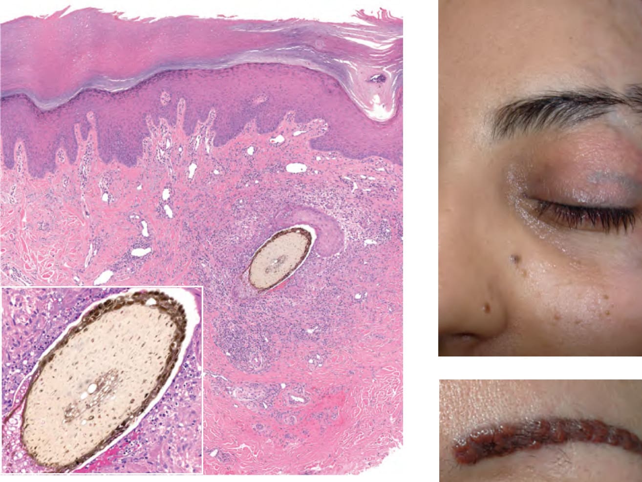
Fig. 94.3 Granulomatous foreign body reaction to implanted organic material in acral volar skin. A plant is the source of the foreign body and the latter is surrounded by a granulomatous reaction as well as neutrophils, lymphocytes, and cellular debris (inset). Superior to the foreign body is a portion of an epidermal cyst due to implantation of epidermal fragments into the dermis. Courtesy Lorenzo Cerroni, MD.
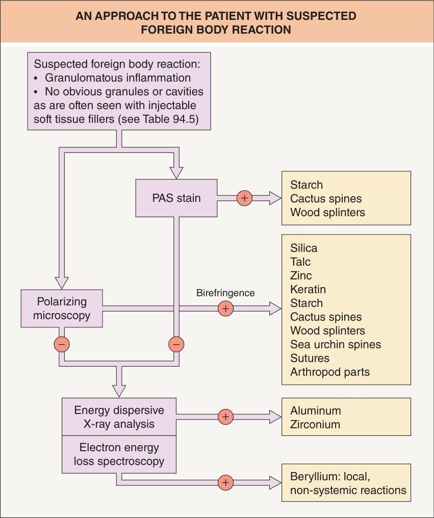
Fig. 94.4 An approach to the patient with suspected foreign body reaction.
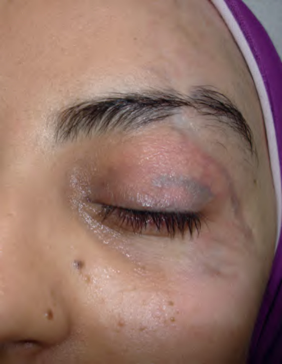
Fig. 94.5 Traumatic tattoo of the eyelid due to asphalt following a motor vehicle accident.
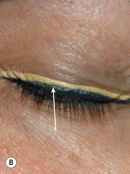
Fig. 94.6 Foreign body reaction to cosmetic tattoos.A Multiple erythematous papules within the eyebrow. B Small papules within black eyeliner (arrow). Courtesy Luis Requena, MD.
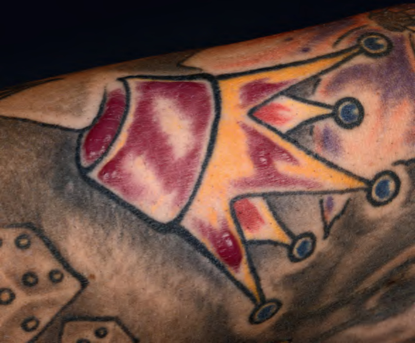
Fig. 94.7 Granulomatous reaction within the red portions of a tattoo. Over the past several years, cinnabar (mercuric sulfide) has been gradually replaced by cadmium selenide (cadmium red), ferric hydrate (sienna), and organic compounds. Courtesy Lorenzo Cerroni, MD.
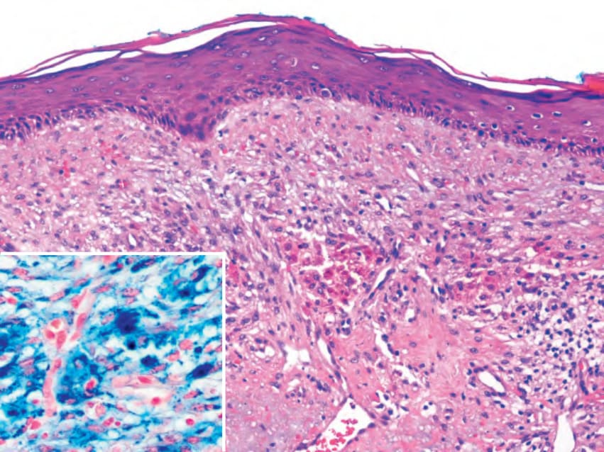
Fig. 94.8 Ferrous subsulfate (Monsel’s solution) – iatrogenic tattoo and granu- lomatous reaction. Coarse granular brown pigment is seen within macrophages and between collagen fibers. The pigment is positive with Perls’ stain (inset). Courtesy Luis Requena, MD.

Table 94.3 Clinical and histopathologic features of foreign body reactions. The order of the entities in this table corresponds to that in the text. Injectable soft tissue fillers are reviewed in Table 94.5. Energy-dispersive X-ray analysis (EDXA) or laser-induced breakdown spectroscopy can be used to detect tattoo inks, silica, talc, zirconium, aluminum, and zinc. iv, intravenous; PAS, periodic acid Schiff.
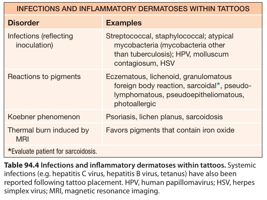
Table 94.4 Infections and inflammatory dermatoses within tattoos. Systemic infections (e.g. hepatitis C virus, hepatitis B virus, tetanus) have also been reported following tattoo placement. HPV, human papillomavirus; HSV, herpes simplex virus; MRI, magnetic resonance imaging.