Silica
Wounds or penetrating injuries accidentally contaminated with silica (silicon dioxide) contained in sand, soil, rocks, and glass may be followed some years later by the formation of a foreign body reaction.
The period between inoculation and development of a reaction may be as long as 25 years. This extended “incubation” period is due to the very slow release of colloidal silica (the only form capable of inciting a reaction) from the large particles of silica introduced into the body. Reactions to silica are thought to be due to a delayed-type hypersensitivity response.
Clinical features
At first, the wound heals, but then papules, nodules, or indurated plaques develop within the scar years later. Multiple disseminated lesions may follow trauma produced by the explosion of land mines and bombs. Patients with sarcoidosis may be at increased risk for immunologic responses to silica.
Pathology
The dermis contains multiple nodular aggregates of granulomas, usually sarcoidal and less often foreign body, separated by strands of connective tissue. Langhans- and foreign body-type giant cells are usually numerous in both types of granuloma. Colorless crystals may be observed within the cytoplasm of the giant cells or lying free in the interstitium. These bodies are birefringent when examined with polarized microscopy.
Differential diagnosis
Granulomatous lesions occurring at the site of an old scar may mimic cutaneous sarcoidosis clinically and histologically. However, the presence of foreign material within a sarcoidal granuloma does not exclude sarcoidosis, given the attraction of sarcoidosis to existing scars. Examination under polarized light will identify silica crystals.
Treatment
Excision of persistent symptomatic lesions is recommended. Spontaneous resolution of silica granulomas, although reported, is exceptional.
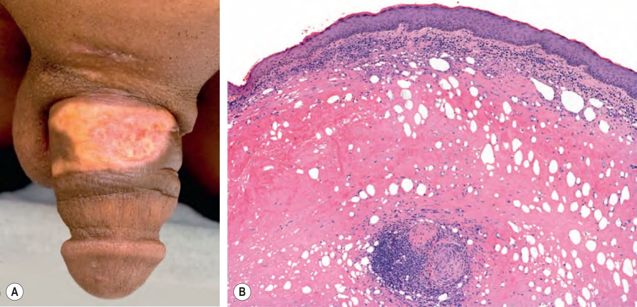
Fig. 94.9 Sclerosing lipogranuloma of the penis.A The penis was injected with paraffin in order to relieve urinary retention following a motorbike accident, and an ulcerated, indurated yellow plaque with telangiectasias developed at the site. B Histologic examination showed sclerosis as well as multiple circular and oval empty spaces within the dermis. Courtesy Glen Foxton, MD and Clare Tait, MD.