PATHOGENESIS
The precise pathogenesis underlying the perforating disorders is unknown. As it is unlikely that dermal connective tissue or hair shafts actively perforate into the epithelium, some have argued that “transepidermal elimination” is a more accurate term. The term “perforating” is embedded in the dermatologic lexicon, however, and is easier to say. Epithelium becomes hyperplastic and eventually surrounds the abnormal connective tissue, just as it appears to do with wood splinters or other foreign bodies. In acquired perforating dermatosis, pruritus probably leads to chronic scratching, which results in epithelial hyperplasia, as in prurigo nodularis. Indeed, prurigo nodules are often admixed with classic perforating lesions. The possibility has been raised that enlargement of a hyperkeratotic plug could lead to penetration through the base of the hyperplastic epidermis, i.e. the perforation is caused by the plug above rather than being due to the abnormal connective tissue below.
Primary perforating diseases may be due to either genetic or acquired abnormalities of collagen or elastic fibers. In EPS, as well as some cases of acquired perforating dermatosis in which perforation of elastic fibers is observed, enhanced expression of elastin receptors has been detected in the epidermis surrounding the elastic material. It has been proposed that scratching exposes keratinocytes to advanced glycation end product (AGE)-modified extracellular matrix proteins, in particular collagen types I and III. This would then lead to terminal differentiation of keratinocytes via the AGE receptor (CD36), followed by upward movement of keratinocytes along with glycated collagen. Increased levels of AGEs have been detected in the skin of patients with acquired perforating dermatoses.
Plasma fibronectin levels are elevated in patients with diabetes mellitus and uremia, and fibronectin is also increased within the skin at sites of transepidermal elimination. This may be significant, since fibronectin plays a role in epithelial cell signaling, locomotion, and differentiation. It binds to type IV collagen (the type found in basement membranes) and to keratinocytes and may incite epithelial proliferation and perforation. In one study of RPC, the investigators demonstrated that the collagen being transepidermally eliminated was type IV collagen. Increased expression of transforming growth factor- β (TGF-β), matrix metalloproteinase-1 (MMP-1), and tissue inhibitor of metalloproteinase-1 (TIMP-1) has been observed in lesional skin of acquired RPC, but this enhanced expression may be a reflection of concurrent wound healing (see Ch. 141).
Other proposed pathomechanisms include abnormal vitamin A or D metabolism, enzyme release from neutrophils, and microangiopathy related to diabetes. Deposition of uric acid, hydroxyapatite, or silicon has also been implicated in the pathogenesis of perforating disorders.
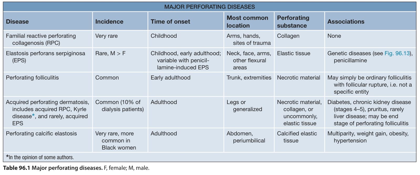
Table 96.1 Major perforating diseases. F, female; M, male.
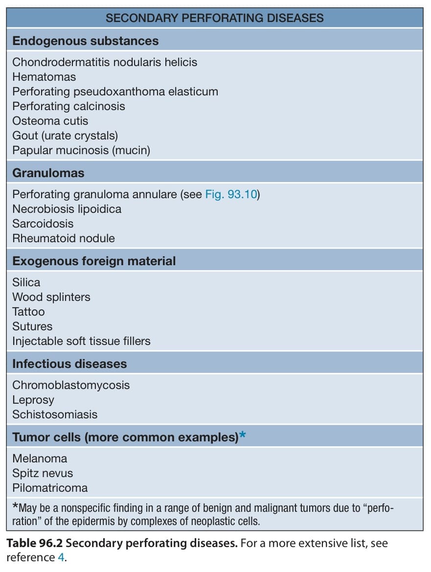
Table 96.2 Secondary perforating diseases. For a more extensive list, see reference 4.