CLINICAL FEATURES
Reactive Perforating Collagenosis
Familial RPC is a rare disorder that begins during childhood. After superficial trauma, patients develop keratotic papules that reach a size
of 5–8 mm over the following 3–4 weeks. The Koebner phenomenon may occur, in which injury to the skin results in the formation of new lesions, often in a linear distribution. Koebnerization is more commonly observed with RPC than with the other perforating disorders, but it has been reported with all of them. Arms and hands are the most common sites of involvement in RPC (Fig. 96.1). The papules tend to spontaneously resolve over 6–10 weeks. Verrucous perforating collagenoma (collagenome perforant verruciformé) is a very rare non-familial variant of RPC in which severe trauma to the skin results in verrucous papules with transepidermal elimination of collagen. Acquired RPC that begins in adulthood usually occurs in association with diabetes mellitus and/or chronic kidney disease (stages 4–5), and these cases are best classified as acquired perforating dermatosis, even though the histopathology is identical to the inherited form of RPC.
Elastosis Perforans Serpiginosa
EPS is a rare disorder whose onset is during childhood or early adulthood. About 40% of cases occur in association with other genetic disorders, including Down syndrome, Ehlers–Danlos syndrome, osteogenesis imperfecta, Marfan syndrome (Fig. 96.2), pseudoxanthoma elasticum (PXE), Rothmund–Thomson syndrome, and acrogeria. In a survey of pediatric dermatologists, the majority performed a detailed history and careful physical examination but did not embark upon an extensive search for these genetic disorders in an otherwise healthy child or adolescent with EPS.
Papules arranged in an annular configuration on the neck.
Very rarely, EPS has been associated with chronic kidney disease (more rarely encountered than the acquired form of RPC) and may overlap with acquired perforating dermatosis. EPS has also been reported following long-term use of penicillamine, usually for 10 or more years. This drug is known to disrupt desmosine cross-links within elastin (see Ch. 97). Because penicillamine is used to treat Wilson disease, the possibility that abnormal copper metabolism plays a pathogenic role in EPS has been raised. However, EPS has also been observed in patients with cystinuria or rheumatoid arthritis who have received penicillamine.
Lesions of EPS are keratotic, 2- to 5-mm papules that tend to be arranged in a serpiginous or annular pattern, most commonly on the lateral neck (Fig. 96.3), but also on the face, arms (Fig. 96.4), or other flexural areas. The rings of papules may reach several centimeters in diameter. Most patients experience no symptoms or only mild pruritus. Like RPC, the lesions may spontaneously resolve, but in general tend to persist, often for several years.
Acquired Perforating Dermatosis and Kyrle Disease
Acquired perforating dermatosis has been used as a catch-all term for those perforating diseases that arise in adults, usually in association with diabetes mellitus and/or the pruritus of chronic kidney disease (stages 4–5). The majority of affected patients with established renal failure
are receiving dialysis, and most of those with diabetes mellitus have associated nephropathy. In individuals requiring dialysis, predictors for the development of acquired perforating dermatosis include diabetes mellitus, reduced circulating levels of intact parathyroid hormone, hypoalbuminemia, and elevated serum levels of high-sensitivity C-reactive protein. This disorder can also occur in conjunction with other causes of pruritus, from insect bites and scabies to lymphoma and hepatobiliary diseases, including primary biliary cholangitis and hepatocellular carcinoma.
Acquired perforating dermatosis can arise from other forms of trauma (not just scratching), including within sites of healing herpes zoster. In addition, acquired perforating dermatosis and perforating folliculitis may develop following exposure to several classes of drugs including TNF inhibitors, epidermal growth factor receptor inhibitors (e.g. gefitinib, panitumumab) and other kinase inhibitors (e.g. nilotinib), dipeptidyl peptidase-4 inhibitors, antivirals (e.g. telaprevir, indinavir), sirolimus, and several monoclonal antibodies (e.g. natalizumab, bevacizumab). Of note, some of these medications more commonly induce an acneiform eruption with disrupted pilosebaceous units rather than a perforating dermatosis. While an association with hypothyroidism and hyperparathyroidism has been reported, most of these patients also had established risk factors (e.g. diabetes, nephropathy). Lastly, in a series of 22 patients with acquired perforating dermatosis, three were healthy and had no known associated illnesses and two were renal transplant recipients.
Acquired perforating dermatosis occurs most commonly on the legs (Fig. 96.5), but generalized or widely scattered papules and nodules can be seen (Figs. 96.6 & 96.7). Extensor surfaces are favored over flexures and the mucous membranes are spared. A clue to the diagnosis is the presence of a central keratotic core, which is sometimes physically removed by the patient. Occasionally, a giant variant is observed,
A Numerous hyperpigmented papules and nodules on the legs in a patient with both chronic kidney disease and diabetes mellitus. B The central portion of these lesions can be keratotic or composed of scale-crust. C Larger nodules in a patient receiving hemodialysis for end-stage kidney disease. B, Courtesy Lorenzo Cerroni, MD; C, Courtesy Frank Samarin, MD.
where lesions may reach 2 cm in diameter. Rarely, secondary infections due to bacteria (e.g. Staphylococcus aureus), atypical mycobacteria (e.g. Mycobacterium abscessus), or fungi (e.g. Mucor spp.) have been reported.
Largely on the basis of histologic findings, patients with acquired perforating dermatosis have been variably designated in the literature as having RPC, EPS, perforating folliculitis, or perforating PXE. However, not only do the pathologic findings vary from lesion to lesion in the same patient, but there are often overlapping features. In addition, since some, but not necessarily all, of these lesions appear to be follicular, and since manipulation of the lesions by patients frequently alters the histologic changes, the term “acquired perforating dermatosis” was proposed to encompass all of these cases. Furthermore, since Kyrle had used the phrase “follicularis et parafollicularis” to emphasize that not all lesions were centered on hair follicles, the eponym Kyrle disease has been used synonymously with acquired perforating dermatosis by some authorities. However, others do not consider Kyrle disease to be an entity or prefer to define it as merely representing the presence of end-stage, excoriated, prurigo nodules of folliculitis.
Perforating Calcific Elastosis
This disorder is characterized by plaques that favor the abdomen, especially the periumbilical region of middle-aged, obese, hypertensive, multiparous Black women (Fig. 96.8). Involvement of the breast in patients with chronic kidney disease has also been observed. The plaques may be verrucous or have keratotic papules at their periphery.
In histologic sections stained with H&E or von Kossa or Verhoeff–van Gieson stains, calcified elastic fibers (identical to those in PXE) are seen undergoing transepidermal elimination. However, affected patients do not manifest other clinical findings of PXE. Similar transepidermal elimination has been observed in patients with dermatitis who applied a commercially available calcium salt water normally used for making bean curd or were exposed to calcium chloride contained in ice-melting salt.
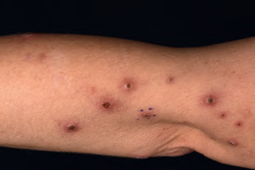
Fig. 96.1 Reactive perforating collagenosis of the arm. Several erythematous papules with a central scale-crust core. Courtesy Lorenzo Cerroni, MD.
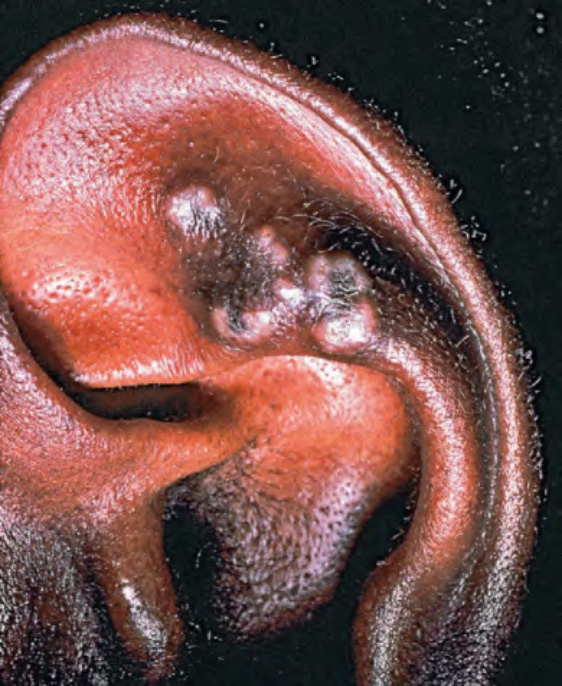
Fig. 96.2 Elastosis perforans serpiginosa in the setting of Marfan syndrome. Annular papules and plaques developed on the ear.
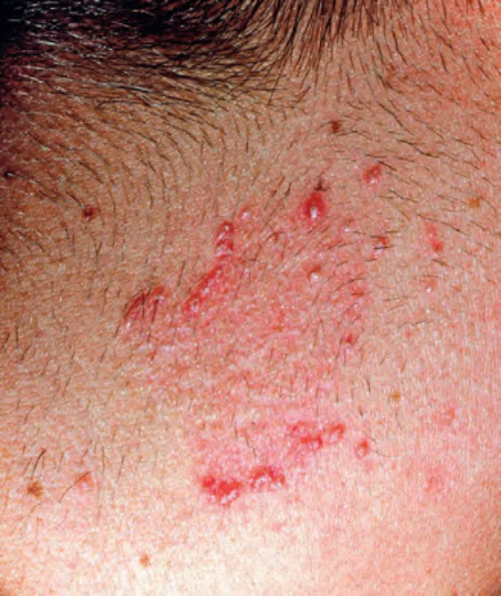
Fig. 96.3 Elastosis perforans serpiginosa.
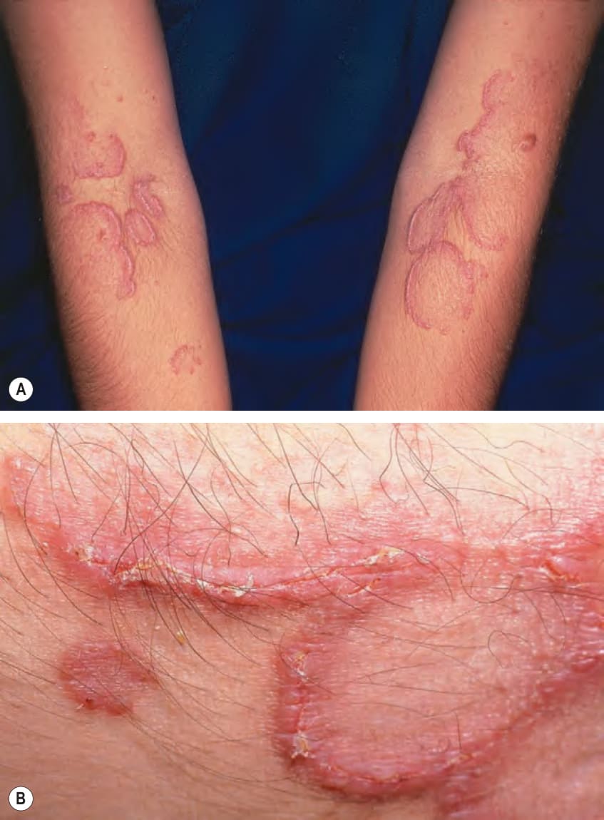
Fig. 96.4 Penicillamine-induced elastosis perforans serpiginosa.A Multiple annular plaques favoring the antecubital fossae. B Closer view with foci of hyperkeratosis at sites of transepidermal elimination.
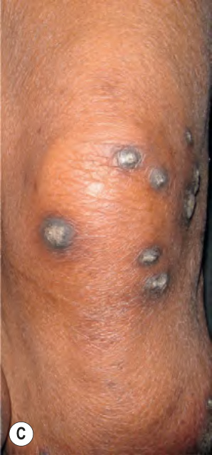
Fig. 96.5 Acquired perforating dermatosis.
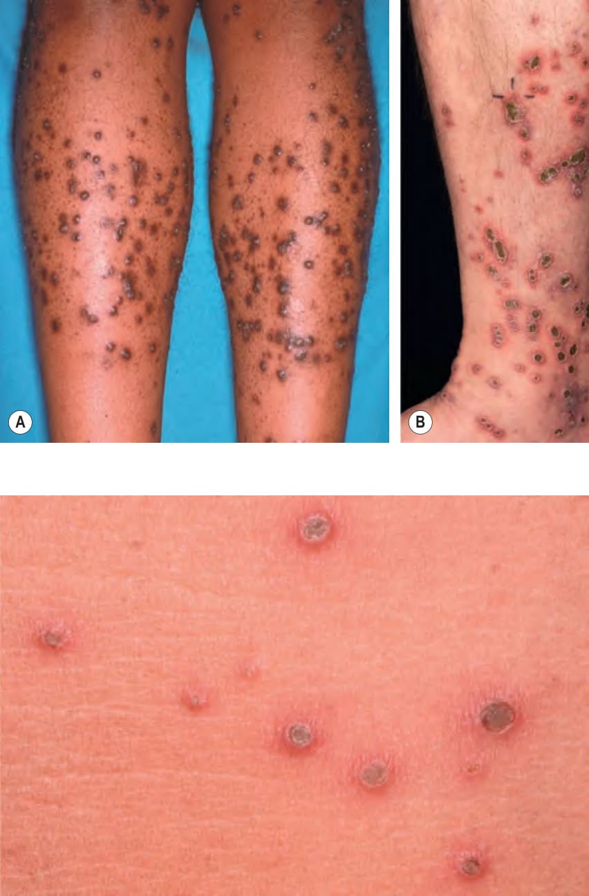
Fig. 96.6 Acquired perforating dermatosis. Erythematous papules with central keratotic cores in a patient with chronic kidney disease. Sometimes the latter are dislodged by the patient. Courtesy Lorenzo Cerroni, MD.
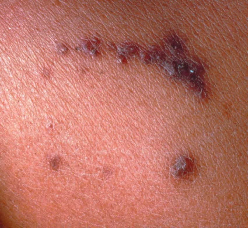
Fig. 96.7 Acquired perforating dermatosis. Note the linear arrangement of the keratotic papules (Koebner phenomenon).
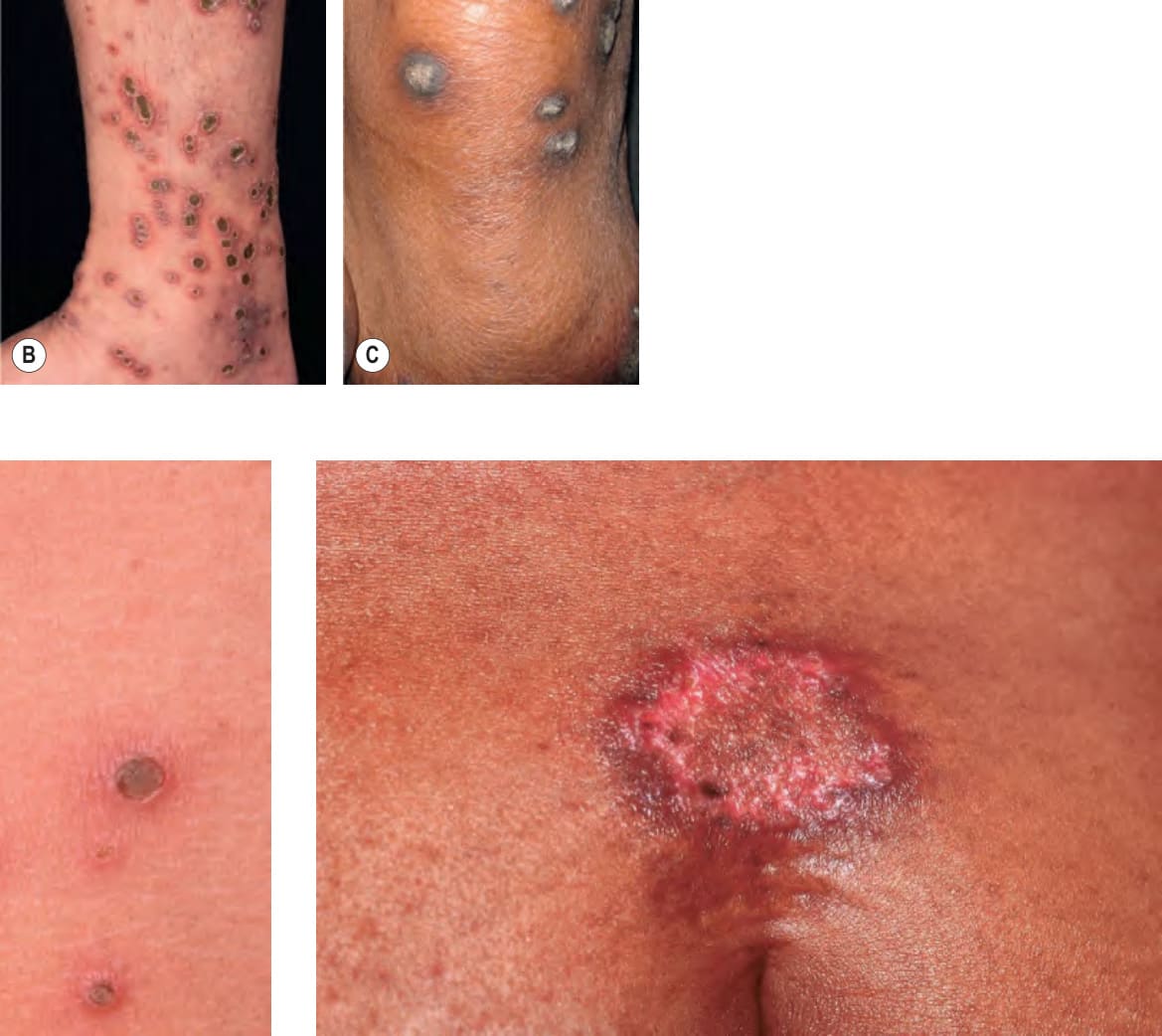
Fig. 96.8 Perforating calcific elastosis. When a biopsy was performed from the elevated edge of this supraumbilical plaque in a multiparous African-American woman, resistance was felt, as well as a grinding sound.