PATHOLOGY
In all the perforating diseases, there is a plug of crusting or hyperkeratosis, with variable parakeratosis, depending upon the stage of the lesion (Fig. 96.9A). As the plug enlarges, epithelial hyperplasia develops, sometimes with dyskeratosis of keratinocytes. At the site of perforation, the epithelial hyperplasia often resembles an elephant snout or crab claw. However, sections from multiple levels through the tissue may need to be examined to find the site of perforation. Some lesions may have aggregates of neutrophils within the dermis, while older lesions, particularly those of EPS, tend to have lymphocytes, macrophages, or multinucleated giant cells in the dermis at the site of perforation.
In RPC, basophilic (degenerative) collagen fibers, some of which are vertically oriented, are seen within the plug or within the epidermis (Figs. 96.9B & 96.10), whereas in EPS, elastic fibers are seen instead (in the same location; Fig. 96.11A). It is important to note, however,
Transepidermal elimination of red collagen fibers through the spinous layer and into the stratum corneum (Verhoeff–van Gieson stain).
that intraepidermal elastic fibers can also be found in other disorders characterized by a hyperplastic epidermis (see below). In RPC, the dermal connective tissue adjacent to the plug appears unremarkable, while in EPS, an increased amount of brightly eosinophilic elastic tissue is often present in the superficial dermis (Fig. 96.11B). The Verhoeff– van Gieson stain is most helpful, because collagen fibers appear red and elastic fibers appear black (Fig. 96.12). However, in EPS, the elastic fibers higher up in the epidermis may lose their ability to stain black. In penicillamine-induced EPS, characteristic “lumpy-bumpy” elastic fibers with lateral buds resembling a “bramble bush” are seen in both lesional and non-lesional skin.
Biopsy findings in acquired perforating dermatosis vary according to the stage of evolution of the lesion. The histology may be identical to childhood RPC or EPS, or to perforating folliculitis. In other cases, it may be less specific, with amorphous degenerated material within the perforations. This material often cannot be clearly identified as collagen or elastic fibers, but sometimes both are present (see Fig. 96.12). A Japanese consensus group recently proposed classification criteria for perforating disorders based on histologic and clinical findings.
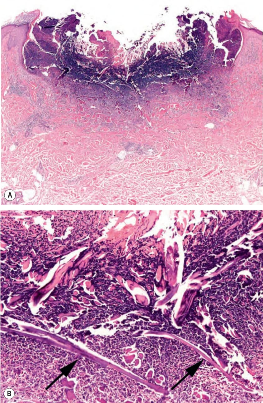
Fig. 96.9 Acquired perforating dermatosis (acquired reactive perforating colla- genosis).A Scanning view of a crusted keratotic plug in a patient whose biopsy showed perforating collagen fibers. B Higher magnification demonstrating collagen fibers (arrows) extending into the crusted plug. Courtesy Lorenzo Cerroni, MD.
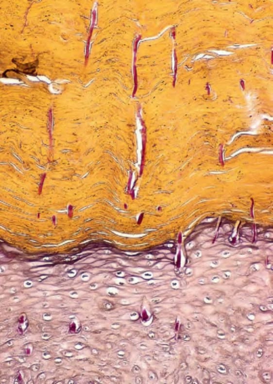
Fig. 96.10 Reactive perforating collagenosis.
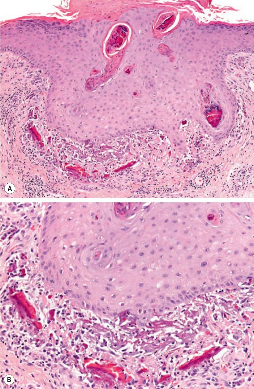
Fig. 96.11 Elastosis perforans serpiginosa.A Hyperplastic epidermis with transepidermal elimination of dermal elastic fibers via channels. Note the surface scale-crust. B Characteristic brightly eosinophilic elastic fibers within the superficial dermis. Courtesy Lorenzo Cerroni, MD.
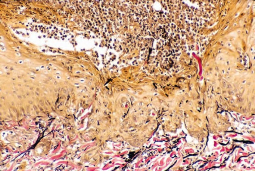
Fig. 96.12 Acquired perforating dermatosis. Transepidermal elimination of both collagen (red) and elastic fibers (black) into a crust with many neutrophils is seen (Verhoeff–van Gieson stain).