HYPERTROPHIC SCARS AND KELOIDS
Key features
Conventional scars are preceded by injury, form within weeks, remain flat or minimally elevated, and are asymptomatic
Hypertrophic scars are raised, firm, and confined to the wound margin. Sometimes they improve spontaneously and tend to have a good response to treatment
Keloids extend beyond the wound margin and are delayed in onset.
They seldom resolve spontaneously and response to treatment is often poor
Introduction
All wounds heal with some degree of scar formation, but the mecha nisms that govern whether the result will be a fine thin scar, a prominent hypertrophic scar, or a tumor-like keloid remain unclear. The latter represent two forms of abnormal wound healing characterized by local fibroblast proliferation and excessive collagen production in response to cutaneous injury. However, their clinical and histopathologic features differ, as well as proposed pathogeneses.
Epidemiology
Hypertrophic scars develop more often than keloids, and keloids have a higher prevalence in individuals with more darkly pigmented skin including Africans, African-Americans, Hispanics, and Asians. In these populations, an incidence ranging from 4%–16% has been reported. Neither hypertrophic scars nor keloids have a sex predilection, and both entities occur most frequently in those 10 to 30 years of age. Individuals in this age group are more often subjected to trauma and their rate of collagen synthesis is higher. Younger skin also possesses greater tension, as compared to older skin which has less elasticity and is more redundant.
Etiology
Proposed etiologic factors include: genetic predisposition; depth, type, and location of the skin injury or wound; degree of tension; local infection or inflammation; and hormonal influences. Possible autosomal dominant inheritance with incomplete clinical penetrance and variable expression has been suggested. A range of skin injuries can lead to abnormal scarring, from lacerations, burns, surgical excisions, and skin piercings to injections (vaccines, tattoo inks) and cutaneous inflammation (e.g. acne vulgaris, insect bites). Moreover, tension is constantly transmitted to the skin from the underlying cartilaginous and bony skeleton, and skin tension can be aggravated by loss of tissue, as occurs with surgical excisions. In addition, hormonal influences have been proposed as an explanation for the appearance of keloids at or after puberty and their resolution following menopause, as well as reports of the onset or enlargement of keloids during pregnancy. Lastly, spontaneous keloids may occur in the absence of any obvious trauma or inflammation, although there is the possibility of unrecognized skin disease (e.g. acne).
Future insights into etiology may come from the genetic disorders in which keloids are an associated feature, including Bethlem myopathy, Rubinstein–Taybi, Warburg–Cinotti and Goeminne syndromes, fronto metaphyseal dysplasia, and other filamin A-related disorders (see Table 95.5). Patients with polyfibromatosis (see below) can also develop keloids.
Pathogenesis
The ability to heal wounds created by injuries, in particular trauma, offers an evolutionary advantage, with associated scarring being less of an issue. However, cutaneous scarring resulting from surgical procedures is viewed as an unwanted consequence. A regenerative response would be preferred, thereby conceivably providing a superior cosmetic result and avoiding the formation of hypertrophic scars and keloids. Notably, in regener ative healing, there is an absence of inflammation in addition to a lack of scarring. In fetuses, cutaneous wounding during the first and second trimesters of gestation does not lead to scarring, and this is thought to reflect a healing process that is occurring via tissue regenerative pathways in a sterile environment and in the absence of inflammation.
Excess scar formation is thought to result from dysregulation during at least one of the three phases of wound healing – inflammatory, prolifer ative, and remodeling/maturation (see Fig. 141.2). Proinflammatory cytokines such as interleukin-6 (IL-6) and IL-8 have been associated with increased scar formation while the anti-inflammatory cytokine IL-10 can lead to a reduction in scar tissue. Several additional factors have been shown to play a role in enhancing fibroblast-related scar formation, including transforming growth factor (TGF)-β, plateletderived growth factor (PDGF), the transcription factor homeobox B13, and components of the Wnt signaling pathway. Greater insights into regulation of crucial components of the remodeling phase, e.g. cessation of granulation tissue formation, will hopefully provide targets for preventative or therapeutic interventions for hypertrophic scarring.
Because keloid formation only occurs naturally in humans, the ability to use animal models is limited. In studies utilizing biopsy specimens of keloids, collagen synthesis by fibroblasts is markedly increased as is the production of TGF-β, a key regulatory factor during the proliferative phase of wound healing. However, different TGF-β isoforms may play distinct and even competing roles in wound healing. Expression of TGF-β1 or TGF-β2 may lead to an increase in scarring, whereas expression of TGF-β3 has been associated with a reduction in scarring. This has led to the introduction of therapies that specifically address these different forms of TGF-β (see Table 98.4).
Clinical Features
Distinguishing clinically between a hypertrophic scar and a keloid is usually straightforward, but sometimes it can be difficult if the lesion is small or of recent onset. The characteristic features of hypertrophic scars and keloids are outlined in Table 98.1. Both have a smooth surface, are firm to palpation, and may be pruritic or painful. Occasionally, they can restrict normal movement of adjacent soft tissues. The color can vary from pink–purple (early lesions) to skin-colored to hypo- or hyper pigmented. While keloids may be more elevated above the skin surface than hypertrophic scars, the key difference is that keloids extend beyond the boundary of the original wound into adjacent normal skin, often with claw-like extensions resembling the pincers of a crab (in Greek, chelè means claw) (Figs. 98.1 & 98.2). In contrast, hypertrophic scars remain confined to the site of the original injury (Fig. 98.3).
Hypertrophic scars usually develop within a few weeks to months after wounding, and they may flatten spontaneously within 1 to 2 years. Most keloids appear within one year of an injury, although the intervening interval may be up to two decades. Of note, keloids do not regress over time. Both hypertrophic scars and keloids favor sites of increased wound tension such as the upper trunk, shoulder, and upper outer arm. However, keloids can also develop in sites such as the earlobe, where there is minimal tension. In addition, keloids can form spontaneously, most often in the midchest region but there is the possi bility of unrecognized skin disease (e.g. acne).
Scar assessment
Scar evaluation ranges from simple scales to technologically advanced devices (e.g. optical coherence tomography) that analyze one or more variables in a reproducible manner. The Vancouver Scar Scale (Table 98.2) and the Patient & Observer Scar Assessment Scale (POSAS) are the most commonly employed assessment tools. These scales have significant inter-patient variation because they score subjective features from color to degree of pain and pruritus. More recently, the Japan Scar Workshop (JSW) published the JSW Scar Scale (JSS), a simple and easy tool for objectively diagnosing keloids and hypertrophic scars
hypertrophic scar.A The keloid has claw-like extensions that extend beyond the original wound margin into adjacent normal skin. B The hypertrophic scar remains confined to the site of the original surgical wound. A, Courtesy Edward Cowen, MD; B, Courtesy Jean L. Bolognia, MD.
based on risk factors of individual patients and sites of involvement. Non-invasive techniques can also be used to more objectively measure scar color, blood flow, transcutaneous oxygen tension, skin hardness, elasticity, and hydration.
Pathology
Distinguishing between a keloid and a hypertrophic scar is important when contemplating treatment, especially more aggressive therapies such as radiation. In some patients, histologic examination is required to make this distinction (Table 98.3). Hypertrophic scars are characterized by an increase in the number of fibroblasts and the density of collagen fibers within the dermis, both of which are oriented parallel to the skin surface. This is accompanied by multiple vertically oriented dermal blood vessels (Fig. 98.4). In keloids, there are whorls and nodules of strik ingly thick, glassy, homogeneous collagen bundles that are composed of densely packed fibrils and are oriented haphazardly throughout the dermis (keloidal collagen) (Fig. 98.5). Early on, there are abundant deposits of fibrillary collagen within the reticular dermis of keloids, while mature lesions often have the characteristic thick sclerotic collagen. In longstanding keloids, there may be a return to the earlier fibrillary pattern.
It is noteworthy that keloidal collagen may be absent in up to 45% of keloids. In scars with no detectable keloidal collagen, histologic features that favor a keloid include: no flattening of the epidermis; a lack of fibrosis within the papillary dermis; a tongue-like advancing edge as the scar tissue extends through the reticular dermis; a horizontal cellular fibrous band within the upper reticular dermis with a sharp demar cation from the normal-appearing papillary and reticular dermis; and prominent fascia-like fibrous bands in the deeper portion of the scar.
Attempts to differentiate hypertrophic scars from keloids via immuno histochemistry has led to conflicting results. In one study, nodules of α-smooth muscle actin (α-SMA)-positive cells (myofibroblasts) were only observed in hypertrophic scars, whereas another investigation found α-SMA expression in both hypertrophic scars (70%) and keloids (45%). Expression of cyclooxygenase (COX)-1 was observed in 100% of keloids, but it was also expressed in ~50% of hypertrophic scars; thus COX-1 expression favored, but did not specifically identify, keloids. Lastly, in vitro differences in fibroblast phenotype, production of extra cellular matrix, and CCL5 secretion have been observed.
Differential Diagnosis
The clinical differential diagnosis of keloids includes the sclerotic form of xanthoma disseminatum, lobomycosis (keloidal blastomycosis; lacaziosis), and the keloidal forms of scleroderma and morphea. Rarely, carcinoma en cuirasse can present as keloidal nodules. Patients with the vascular type of Ehlers–Danlos syndrome may occasionally develop keloidal plaques on their lower extremities, and spontaneous formation of keloids can be a clinical clue to other rare inherited syndromes such as Bethlem myopathy, Rubenstein–Taybi syndrome, and Goeminne syndrome (see above).
Histologically, keloids and hypertrophic scars need to be differentiated from spindle cell tumors, primarily dermatofibroma (DF), dermato fibrosarcoma protuberans (DFSP), dermal fibromatoses, desmoplastic melanoma, and the scar-like variant of squamous cell carcinoma (SCC). Immunostaining for CD34 and factor XIIIa, which is usually positive in DFSP and DF, respectively, is negative in keloids and hyper trophic scars. While there is minimal or no immunostaining for S100 protein in scars, desmoplastic melanomas usually demonstrate strong expression of this marker. Finally, the unusual scar-like variant of SCC may only have focal keratin expression, and a complete panel of keratin stains (to increase sensitivity) may be required for its detection.
Treatment
The management of keloids and hypertrophic scars continues to challenge clinicians, and there is no universally accepted treatment algorithm. Potential evidence-based treatments are outlined in Table 98.4. Prevention remains the best strategy in predisposed patients, including avoidance of nonessential surgery in high-risk anatomic sites and attention to postsurgical wound care. International guidelines recommend use of silicone gels and sheets in the treatment of both hypertrophic scars and keloids. While potent topical corticosteroids
Shading indicates differences.
Adapted from Sullivan T, Smith J, Kermode J, et al. Rating the burn scar. J Burn Care Rehabil 1990;11:256–60.
are sometimes used to address pruritus, intralesional triamcinolone remains a commonly employed first-line therapy. Incremental increases in triamcinolone concentration may be used to balance flattening effects against the risk of atrophy. An alternative, especially for needle-averse individuals, is clobetasol propionate 0.05% cream under silicone dressing occlusion.
Intralesional or topical corticosteroids may be combined with intra lesional 5-fluorouracil. For hypertrophic scars and keloids in patients with darker skin color who are at risk for steroid-induced hypopigmen tation, intralesional bleomycin and intralesional triamcinolone have been shown to lead to comparable improvement. By reducing muscle tension during wound healing, intralesional botulinum toxin type A can minimize scar formation and reduce associated symptoms; possible explanations include pausing of the fibroblast cell cycle and changes in TGF-β1 expression. Based upon case reports, dupilumab can reduce associated pruritus and may lead to an improvement in appearance34a.
Surgical revision of hypertrophic scars and keloids can quickly relieve symptoms, but the recurrence rate is quite high. Cryosurgery has been used successfully and is often combined with intralesional triamcin olone. Shave excision followed by contact cryosurgery has also been employed. Pulsed dye laser therapy has become popular as a modality for postsurgical scar reduction and is associated with few side effects. The use of lower cumulative doses has improved the safety of adjuvant
postoperative radiation therapy while still maintaining efficacy (see Table 98.4 & Ch. 139).
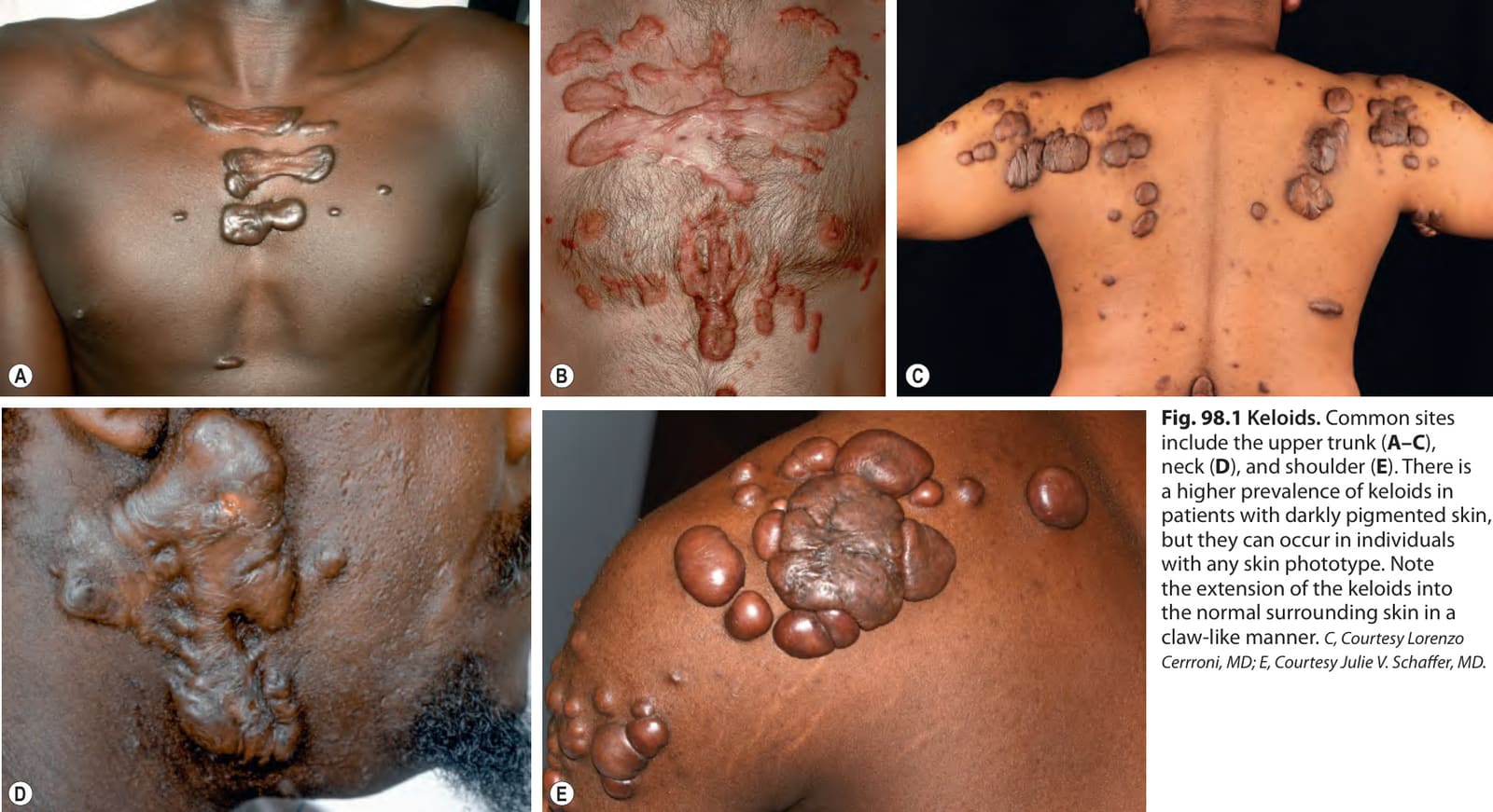
Fig. 98.1 Keloids. Common sites include the upper trunk (A–C), neck (D), and shoulder (E). There is a higher prevalence of keloids in patients with darkly pigmented skin, but they can occur in individuals with any skin phototype. Note the extension of the keloids into the normal surrounding skin in a claw-like manner. C, Courtesy Lorenzo Cerrroni, MD; E, Courtesy Julie V. Schaffer, MD.
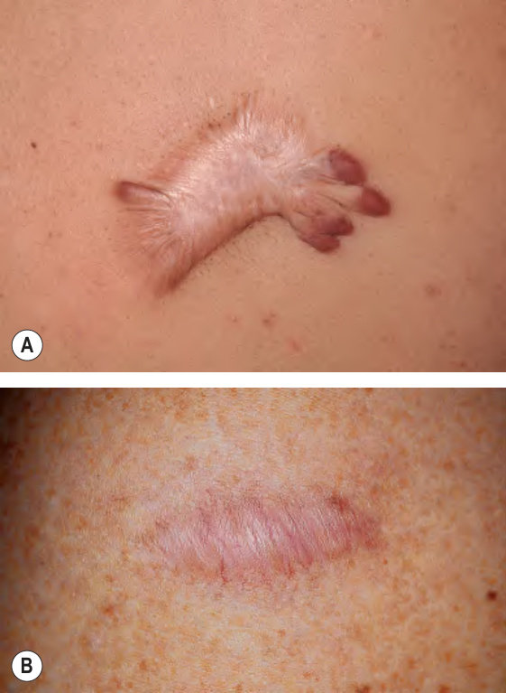
Fig. 98.2 Comparison of a keloid and a
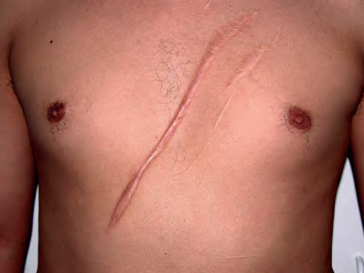
Fig. 98.3 Hypertrophic scar at the site of a knife injury.
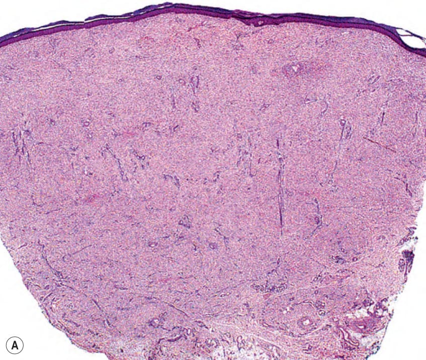
Fig. 98.4 Hypertrophic scar – histopathologic features.A Increased density of collagen fibers within the dermis and vertically oriented blood vessels. B Increase in the number of fibroblasts and density of collagen fibers, both of which are oriented horizontally (parallel to the epidermis). The central blood vessel is oriented vertically and there is a sparse perivascular inflammatory infiltrate.
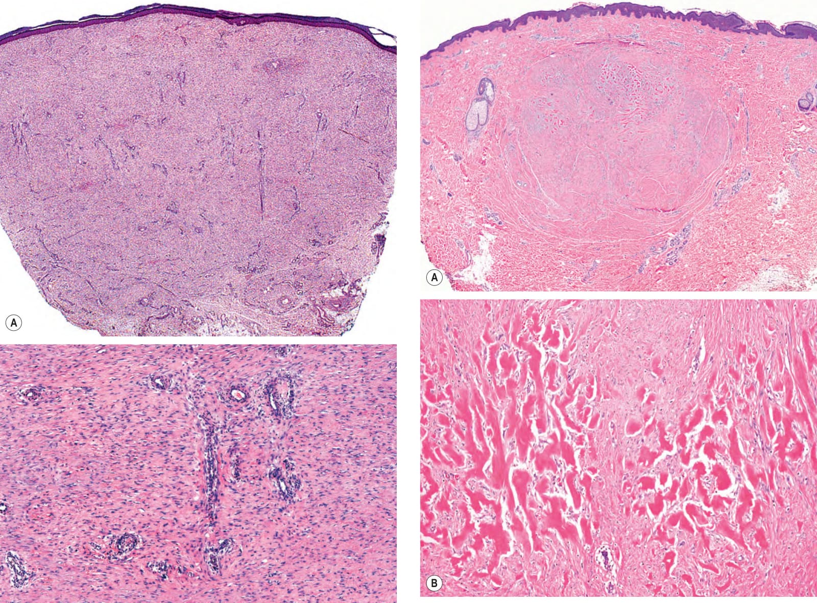
Fig. 98.5 Keloid – histopathologic features.A Nodules of thick collagen bundles are oriented haphazardly within the dermis (keloidal collagen). There is neither flattening of the epidermis nor involvement of the papillary dermis, and a sharp demarcation is seen between the normal-appearing papillary and reticular dermis and the nodules. B Strikingly thick, glassy, homogeneous collagen bundles composed of multiple, densely packed fibrils.
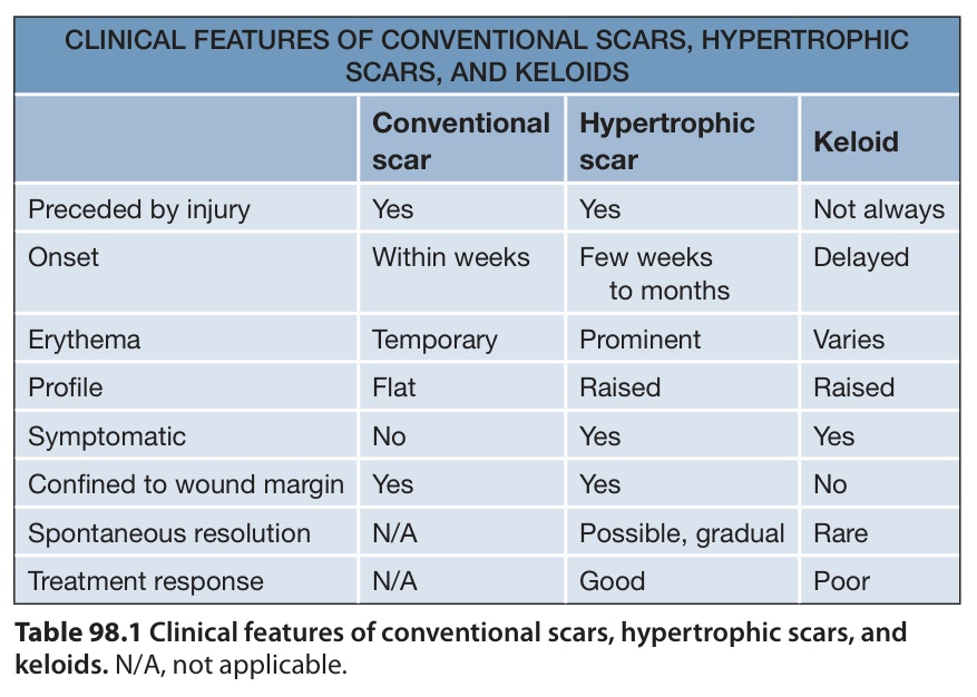
Table 98.1 Clinical features of conventional scars, hypertrophic scars, and keloids. N/A, not applicable.

Table 98.2 Vancouver Scar Scale.
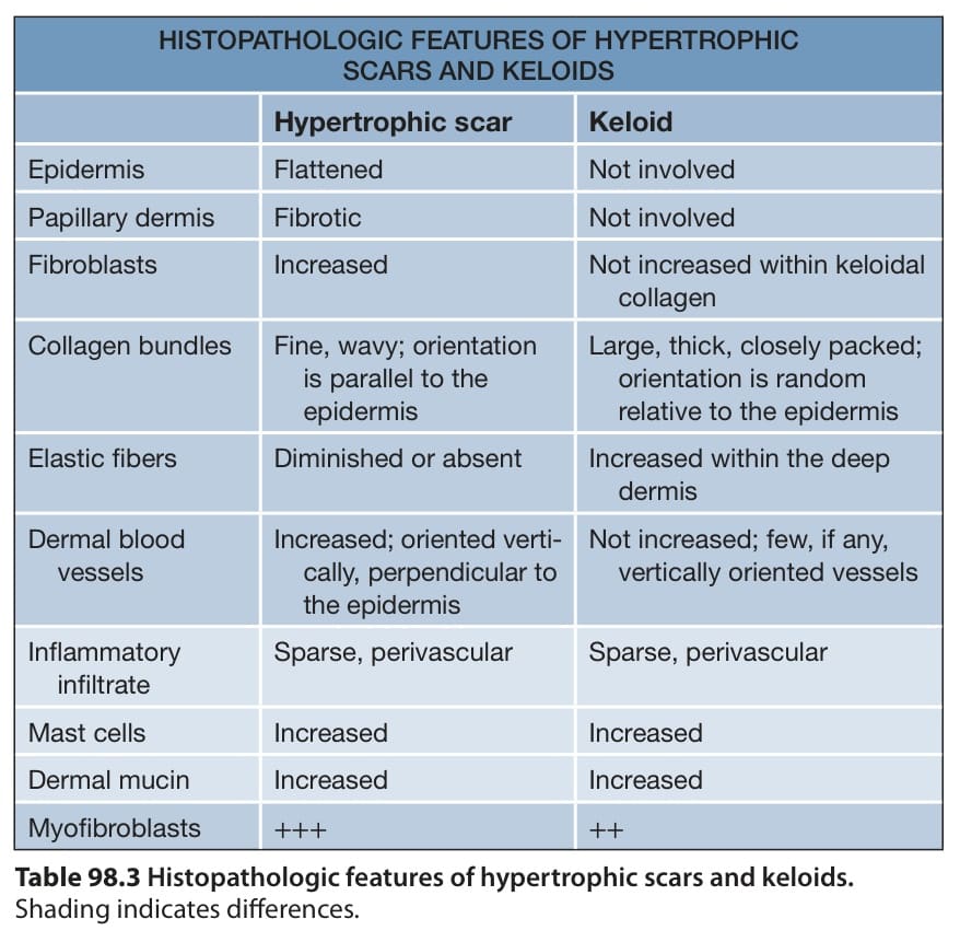
Table 98.3 Histopathologic features of hypertrophic scars and keloids.

Table 98.4 Treatment options for hypertrophic scars and keloids. There are also reports of the use of topical retinoic acid, calcineurin inhibitors, imiquimod (deemed ineffective in controlled trials), and tamoxifen; mitomycin C (topical or intralesional); and systemic methotrexate, dupilumab, and calcium channel blockers. Treatments reserved for the treatment of keloids are shaded mid-blue and experimental treatments are shaded dark blue. Key to evidence-based support: (1) prospective controlled trial; (2) retrospective study or large case series; (3) small case series or individual case reports. TGF, transforming growth factor.