DUPUYTREN DISEASE
Synonyms: Dupuytren’s contracture Palmar fibromatosis Dupuytren’s diathesis
Key features
Thickening of palmar and digital fascia
Progressive flexion contracture of affected digits
Myofibroblast proliferation followed by excess collagen synthesis
Treatment includes injections of collagenase, percutaneous needle fasciotomy, and surgical fasciotomy/fasciectomy
History
Henry Cline described thickening of the palmar fascia in 1777 and proposed palmar fasciotomy as a cure. In 1831, Guillaume Dupuytren characterized and popularized this entity.
Adapted from Tziotzios C, et al. J Am Acad Dermatol 2012;66:13–24.
Epidemiology
Dupuytren disease is common in White people of northern European ancestry, with a prevalence in Western countries that ranges from 1%–30%; it is rarer in patients with darker skin phototypes. The incidence of Dupuytren disease increases with age and it is most common in middle-aged and older men. Familial associations suggest an autosomal dominant inheritance, but sporadic cases are common. The lesion is associated with other fibromatoses (e.g. plantar fibroma tosis, polyfibromatosis; see Ch. 116) as well as diabetes mellitus, liver disease including alcoholic cirrhosis, and perhaps epilepsy, manual labor, and cigarette smoking.
Vemurafenib has been linked to new-onset or worsening of Dupuytren disease and plantar fascial fibromatosis. To date, there has been one report of the association of topical timolol with the development of Dupuytren disease and penile fibromatosis.
Pathogenesis
Fibrogenic cytokines (e.g. TGF-β1, TGF-β2) are thought to play a role due to their ability to induce collagen production, proliferation of fibroblasts, and the differentiation of fibroblasts into myofibroblasts. Production of tumor necrosis factor (TNF) by M2 macrophages and mast cells in early-stage Dupuytren disease is also thought to activate myofibroblasts, leading to and/or sustaining fibrosis. In genome-wide assays of fibroblasts from Dupuytren disease, the differential expression patterns included genes that encode proteins involved in the extracel lular matrix (e.g. collagen), fibrosis (e.g. follistatin), tissue remodeling (e.g. collagenases, matrix metalloproteinases), cellular movement (e.g. kinesins), and signaling pathways (e.g. STAT1, Wnt2), as well as growth factors (e.g. fibroblast growth factor 9). Activation of the Wnt signaling pathway appears to play a key role as it does in scar formation (see above).
Clinical Features
Patients often present late in the course of their disease with flexion contractures of the affected digits. Lesions usually have an ulnar distri bution, and the ring finger is most commonly affected (Fig. 98.6). The disorder begins with a nodule in the fascia of the palm, which grows to produce a cord which then contracts, producing a flexion contracture of the metacarpophalangeal and proximal interphalangeal joints. Complications of Dupuytren disease include nerve injury, loss of joint mobility, and development of complex regional pain syndrome (also referred to as reflex sympathetic dystrophy).
Pathology
Histologically, the nodular lesion consists of a proliferation of myofibro blasts, which align along lines of tension. The initial stages are more cellular and there are some mitotic figures; the cells may express α-SMA, indicative of myofibroblastic differentiation. Thicker collagen, fewer myofibroblasts, and scarce to absent mitotic figures characterize later stages.
Differential Diagnosis
Although usually the diagnosis is straightforward, one must consider a ganglion cyst, giant cell tumor of the tendon sheath, soft tissue sarcoma (e.g. epithelioid sarcoma), callus, and tenosynovitis. An ultra sonographic hypoechoic mass superficial to a flexor tendon of the third, fourth, or fifth digits at the level of the distal metacarpal, where the length of the lesion is greater than its width, is typical for palmar fibro matosis and helps to exclude other diagnoses.
A rare case of lung cancer metastasis simulating Dupuytren disease has been reported, and in injection drug users, flexion digital contrac tures (camptodactyly) due to repeated vascular injury and infection of the digits may mimic Dupuytren disease. Flexion contractures of the hand, along with multiple tender nodules, can develop in the rare paraneoplastic palmar fasciitis and polyarthritis syndrome.
Treatment
Although surgical fasciotomy/fasciectomy to release the joint contracture is the mainstay of treatment, minimally invasive procedures such as percutaneous needle fasciotomy and enzymatic fasciotomy with injectable collagenase (from Clostridium histolyticum) are increas ingly utilized despite higher recurrence rates. Patients receive up to three monthly injections of collagenase per cord, followed by passive finger extension 1–3 days later. Complications include bruising, tendon rupture, ligament or nerve injury, and anaphylaxis. Although some authors have advocated the use of radiotherapy during the early stage of Dupuytren disease, a systematic review found limited evidence to support this approach.
A risk score determined by whole genome sequencing may predict postsurgical recurrence, while ultrasound findings (e.g. mixed echogenicity, nodular structure) may predict recurrence after minimally invasive treatments. Additional possible treat ments include intralesional triamcinolone, blue light irradiation (to prevent relapse), correction of vitamin D deficiency, and use of inhibitors of TGF-β1, TGF-β2, TNF and one of its receptors TNFR2, or IL-33. New enzymatic approaches are also in development.
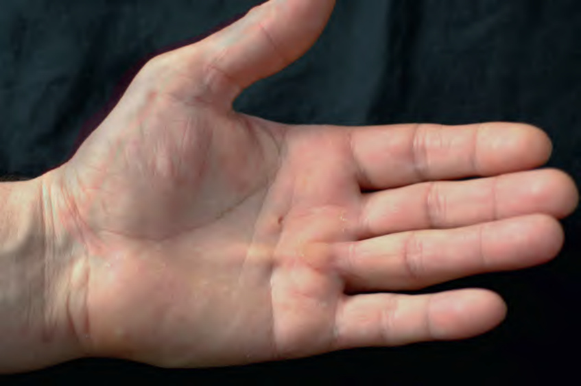
Fig. 98.6 Dupuytren disease. Fibrotic nodule at the base of the fourth finger with an obvious cord extending proximally. In addition to palpation, fibrotic cords can be accentuated by extension of the digit. Eventually, flexion contractures develop.
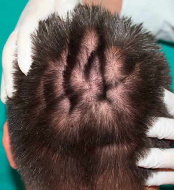
Fig. 98.7 Cutis verticis gyrata. Cerebriform folding of the skin of the scalp.

Table 98.4 Treatment options for hypertrophic scars and keloids. There are also reports of the use of topical retinoic acid, calcineurin inhibitors, imiquimod (deemed ineffective in controlled trials), and tamoxifen; mitomycin C (topical or intralesional); and systemic methotrexate, dupilumab, and calcium channel blockers. Treatments reserved for the treatment of keloids are shaded mid-blue and experimental treatments are shaded dark blue. Key to evidence-based support: (1) prospective controlled trial; (2) retrospective study or large case series; (3) small case series or individual case reports. TGF, transforming growth factor.