HYALINE FIBROMATOSIS SYNDROME (JUVENILE HYALINE FIBROMATOSIS AND INFANTILE SYSTEMIC HYALINOSIS)
Synonyms: Murray–Puretic–Drescher syndrome Fibromatosis hyalinica multiplex juvenilis (Juvenile) systemic hyalinosis
Key features
Rare autosomal recessive disorder with variable phenotypic expression
Usually presents during infancy or early childhood
Papulonodules that favor the ears, scalp, neck, hands, and periorificial areas plus gingival hypertrophy and flexion contractures of large joints
Mutations in the gene that encodes anthrax toxin receptor 2 (ANTXR2)
Treatment consists of excision of nodules and gingivae as needed, but lesions often recur
Introduction
Hyaline fibromatosis syndrome (HFS) is a rare autosomal recessive genodermatosis characterized by hyaline accumulation within the dermis. HFS encompasses both juvenile hyaline fibromatosis (JHF) and infantile systemic hyalinosis (ISH). These two disorders are allelic, i.e. they are due to mutations in the same gene, but differ in severity and visceral involvement (Table 98.6).
History
In 1873, Murray described a condition which he termed “molluscum fibrosum”. In 1972, Kitano and colleagues renamed the disorder “juvenile hyaline fibromatosis”.
Epidemiology
HFS is a rare disorder that presents during infancy or early childhood.
Pathogenesis
Mutations are present in the gene that encodes anthrax toxin receptor 2 (ANTXR2), also referred to as capillary morphogenesis protein-2 (CMG2). ANTXR2 is a transmembrane protein that binds to laminins and types IV and VI collagen (as well as the anthrax toxin). In the skin it is thought to play a role in cell–matrix interactions and basement membrane integrity. Because ANTXR2/CMG2 is thought to target collagen VI for degradation, its dysfunction leads to an accumulation of collagen VI and a subsequent sequestration of inactive matrix metalloproteinases by excess collagen VI, thus leading to fibrosis. As noted previously, mutations in ANTXR2 can lead to either mild or severe forms of HFS. Missense and other in-frame mutations of the cytoplasmic domain tend to cause a phenotype with a better prognosis (JHF), whereas truncating and missense mutations of the extracel lular protein-binding domain lead to a more severe phenotype (ISH). Because ANTXR2 is not expressed in the brain, mental development is not impaired.
Clinical Features
Initially, firm papules and nodules develop on the scalp, ears, neck, and face (particularly perinasal and perioral regions) as well as the perianal area (Fig. 98.9); papulonodules also favor the hands. Patients often suffer severe physical limitations from joint contractures and tumors. Additional clinical features are outlined in Table 98.6. Milder phenotype and later onset has been observed in females.
Pathology
Ill-defined nodules composed of dense, homogeneous, hyaline (PAS-positive) collagen are seen within the dermis and, in larger lesions, the subcutaneous tissue (Fig. 98.10). Mononuclear cells and CD68+, osteoclast-like giant cells may be present in a perivascular location in addition to perivascular calcification and eosinophilic globules. Over time, the lesions tend to become more paucicellular, and calcospherites or larger areas of calcification are frequently seen (Fig. 98.10, inset).
Differential Diagnosis
HFS needs to be distinguished from the autosomal recessive disorders MONA (multicentric osteolysis, nodulosis, and arthropathy) and Winchester syndrome which are due to mutations in genes that encode matrix metalloproteinases. Both can have joint contractures, coarse facies, thickened skin and gingival hypertrophy, with painful nodules, especially of the soles, in the former. Histologically, both disorders feature a proliferation of fibroblasts and thickened collagen bundles
extending from the deep dermis to the hypodermis. Other genoderma toses to consider include Farber lipogranulomatosis, lipoid proteinosis, infantile myofibromatosis, terminal osseous dysplasia, and the rarer diffuse variant of stiff skin syndrome that is due to fibrillin-1 mutations and inherited in an autosomal dominant pattern.
Treatment
Treatment options are limited. Surgical excision of nodules and gingival hypertrophy and release of contractures can be performed, but lesions often recur. Diode laser therapy improved skin and mucosal lesions in one patient, but these recurred. Physiotherapy may slow the progression of joint contractures. Injections of corticosteroids into fibromas have met with limited success. Subcutaneous interferon-α-2b therapy improved the skin lesions, gingival hypertrophy, and joint mobility in a patient
with JHF. Based on in vitro results, proteasome inhibitors may represent a future therapy for HFS patients with ectodomain-related mutations in ANTXR2.
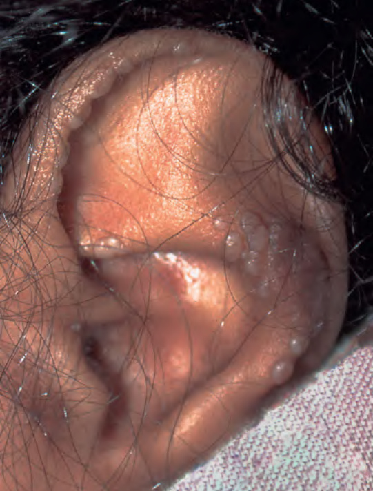
Fig. 98.9 Juvenile hyaline fibromatosis. Firm pearly papules favor the ears.
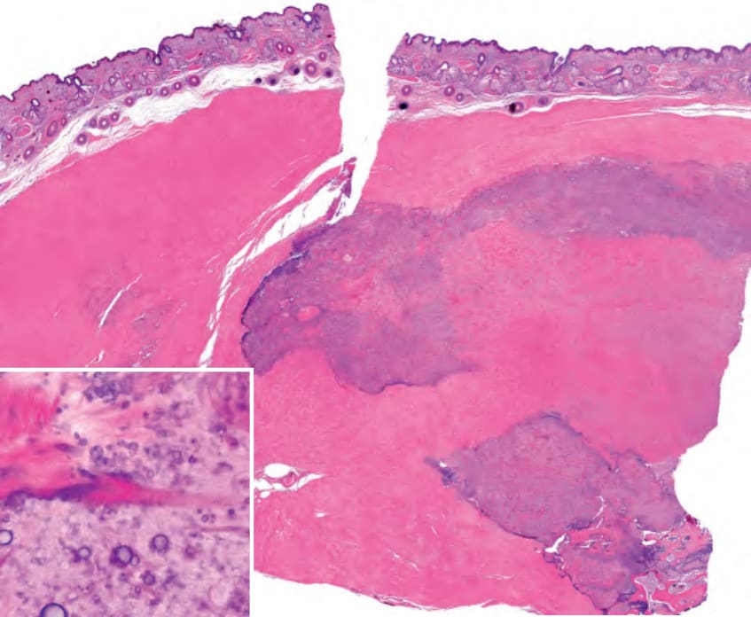
Fig. 98.10 Juvenile hyaline fibromatosis – histopathologic features. Abundant, dense, homogeneous, hyaline (PAS-positive) collagen within the lower dermis and subcutis. Round basophilic calcospherites are also present (inset).
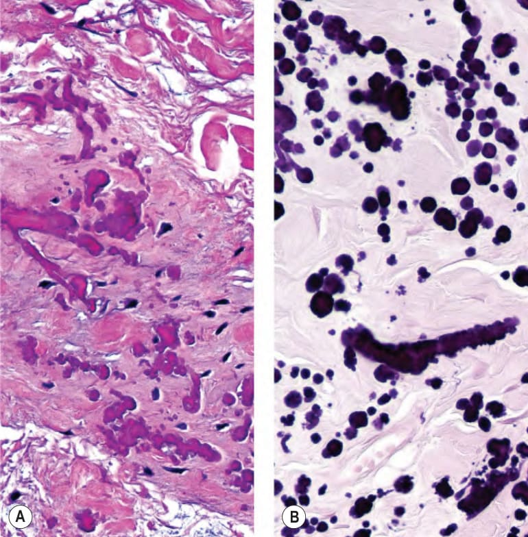
Fig. 98.11 Elastofibroma dorsi – histopathologic features.A Irregular, partially fragmented elastic fibers admixed with swollen collagen bundles. B An elastic tissue stain accentuates the fragmented fibers.
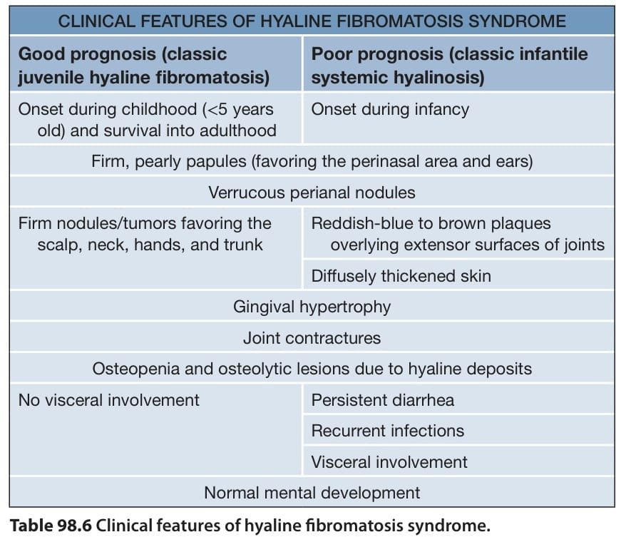
Table 98.6 Clinical features of hyaline fibromatosis syndrome.
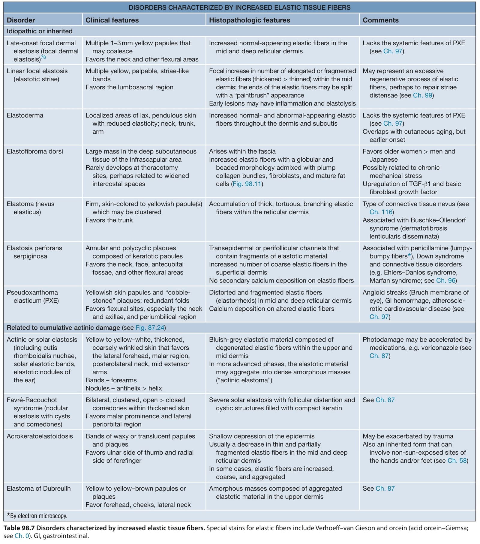
Table 98.7 Disorders characterized by increased elastic tissue fibers. Special stains for elastic fibers include Verhoeff–van Gieson and orcein (acid orcein–Giemsa; see Ch. 0). GI, gastrointestinal.