MID-DERMAL ELASTOLYSIS
Key features
Uncommon disorder with areas of fine wrinkling
Usually affects White middle-aged women
Selective loss of elastic tissue in the mid dermis
Introduction
Mid-dermal elastolysis (MDE) is a rare, acquired disorder of elastic tissue. Clinically, it is usually characterized by diffuse fine wrinkling, most often located on the trunk, shoulders, neck, and arms. Histologically, a clear band of elastolysis with loss of elastic fibers is present in the mid dermis.
History
In 1977, Shelley and Wood reported the first case of “wrinkles due to idiopathic loss of mid-dermal elastic tissue”. Their patient, a 42-year-old woman, had circumscribed areas of fine wrinkles that gave her an inappropriately aged appearance.
Epidemiology
To date, around 100 cases have been reported in the literature. The vast majority of patients are White women between the ages of 30 and 50 years.
Pathogenesis
The cause of the acquired elastic tissue degeneration in MDE has yet to be determined. Exposure to UV light, including natural sunlight, UVA (tanning salon), and narrowband UVB phototherapy, was thought to be a contributing factor in MDE, but this remains unclear. Other hypotheses include defects in the synthesis of elastic fibers, autoimmunity against elastic fibers, and damage to elastic fibers via the release of elastase by inflammatory cells or fibroblasts. Of note, MDE has been observed in the setting of immune reconstitution inflammatory syndrome (IRIS). An imbalance between matrix metalloproteinases (MMPs) and tissue inhibitors of metalloproteinases (TIMPs) may also be playing a role, in addition to CD34+ dendritic fibroblasts.
A decrease in lysyl oxidase-like 2 (LOXL2) expression, potentially affecting elastin renewal, has been observed in MDE. There is also a decrease in dermal fibulin-4 and -5, pointing to altered re-assembly of elastic fibers in MDE, and not simply enhanced elastolysis. In sum, the pathogenesis of MDE is likely multifactorial, with genetic predisposition, chronic inflammation, UV irradiation, and/or (auto)immune processes as contributing factors.
Clinical Features
Patients can have well-circumscribed to large diffuse areas of fine wrinkling (Fig. 99.1), usually in a symmetric distribution (type I). The wrinkles themselves tend to follow cleavage lines. Discrete perifollicular papules are seen in some patients (type II), with the site of the central hair follicle being indented. Occasionally, erythematous patches, telangiectasias, and reticulated erythema may be present (type III). Although in the majority of patients there is no history of a prior inflammatory dermatosis, some patients report previous mild to moderate erythema and, rarely, elastolysis is preceded by urticarial lesions or granuloma annulare.
Sites of predilection are the trunk, lateral neck, and upper extremities. Once the patches of wrinkling have appeared, they usually remain stationary. They are asymptomatic and give the skin a prematurely aged appearance. The affected areas usually have normal pigmentation and no associated scaling, induration, or herniation. There is typically no family history of similar lesions and unless linked to an autoimmune disorder, no systemic involvement. MDE can lead to significant cosmetic concern.
While diagnosis is usually confirmed via histologic examination of involved skin, non-invasive imaging techniques such as optical coherence microscopy or high-frequency ultrasound may also prove helpful.
Pathology
The epidermis is normal in appearance and, occasionally, a mild perivascular infiltrate is noted in the dermis. Elastic tissue stains, such as Verhoeff–van Gieson or Weigert’s stain, reveal a selective loss of elastic fibers in the mid dermis (Fig. 99.2). There is preservation of normal elastic tissue in the superficial papillary dermis above, in the reticular dermis below, and along adjacent hair follicles. The preservation of elastic tissue around the hair follicles explains the perifollicular papules observed in some patients.
By electron microscopy, phagocytosis of normal as well as degenerated elastic fiber tissue by macrophages has been described. Elastophagocytosis by macrophages can also be observed by routine histology.
Differential Diagnosis
MDE must be differentiated from other disorders of elastic tissue such as anetoderma, pseudoxanthoma elasticum (PXE), PXE-like papillary dermal elastolysis, elastoderma, and cutis laxa, especially the acquired form (Table 99.1).
Clinically, anetoderma is characterized by soft macules and papules that herniate upon palpation, as opposed to diffuse wrinkling. Histologically, elastolysis can occur in the papillary and/or mid-reticular dermis in anetoderma (see below). Patients with heritable forms of generalized cutis laxa have loose, redundant skin, hanging in folds, and histologic examination shows reduced and fragmented elastic fibers, usually within the entire dermis. Acquired cutis laxa, both generalized and acral, can be a manifestation of an underlying paraproteinemia, with binding of immunoglobulins to elastic fibers (see Table 97.8).
Less often, MDE is confused with solar elastosis or the perifollicular elastolysis that is usually seen on the trunk in association with acne
vulgaris. Solar elastosis differs clinically by its onset in an older age group, restriction to only sun-exposed areas, yellow color, and coarser wrinkling and differs histologically by hyperplasia of abnormal elastic fibers and basophilic degeneration of the collagen in the papillary dermis. Perifollicular elastolysis leads to a selective and almost complete loss of the elastic fibers that surround hair follicles, compared with preser vation of elastic fibers around follicles in MDE.
Treatment
Currently, there is no effective treatment for MDE. Sunscreens, colchicine, chloroquine, vitamin E, and topical agents (retinoic acid, corticosteroids) have been tried without success. Topical soybean extract and eicosapentanoic acid are potential future therapies and oral mycophenolate mofetil may be beneficial.
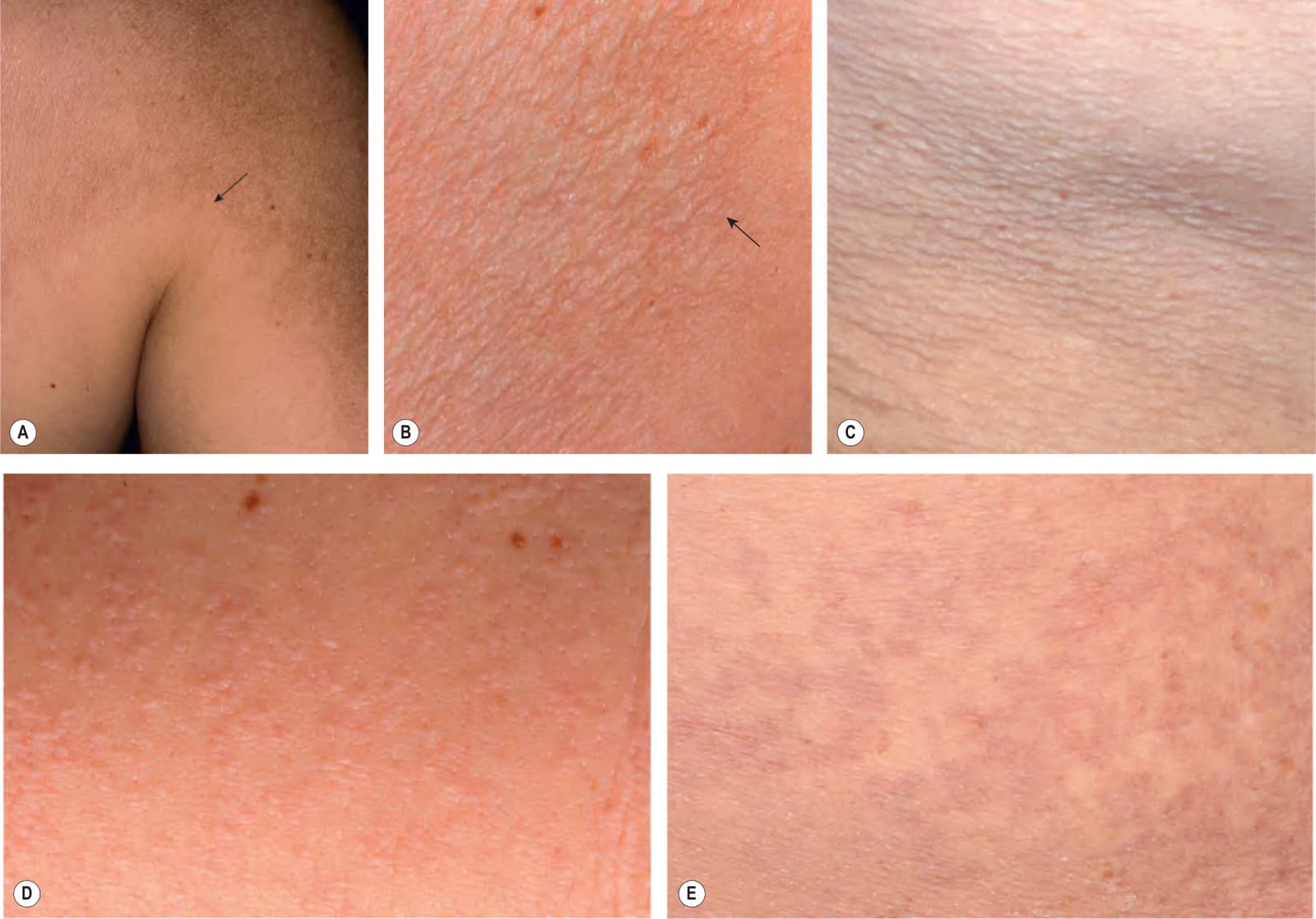
Fig. 99.1 Mid-dermal elastolysis.A, B Type I – large diffuse patch as well as reticulated areas of fine wrinkling on the upper trunk and proximal arm. The junction of involved and uninvolved skin is marked with an arrow. C Types I and II – both circumscribed areas of wrinkling and follicular papules are present. D Type II – multiple skin-colored follicular papules. E Type III – primarily reticulated erythema. A, Courtesy Judit Stenn, MD; B, Courtesy Carlo F. Tomasini, MD; C–E, Courtesy Lorenzo Cerroni, MD.
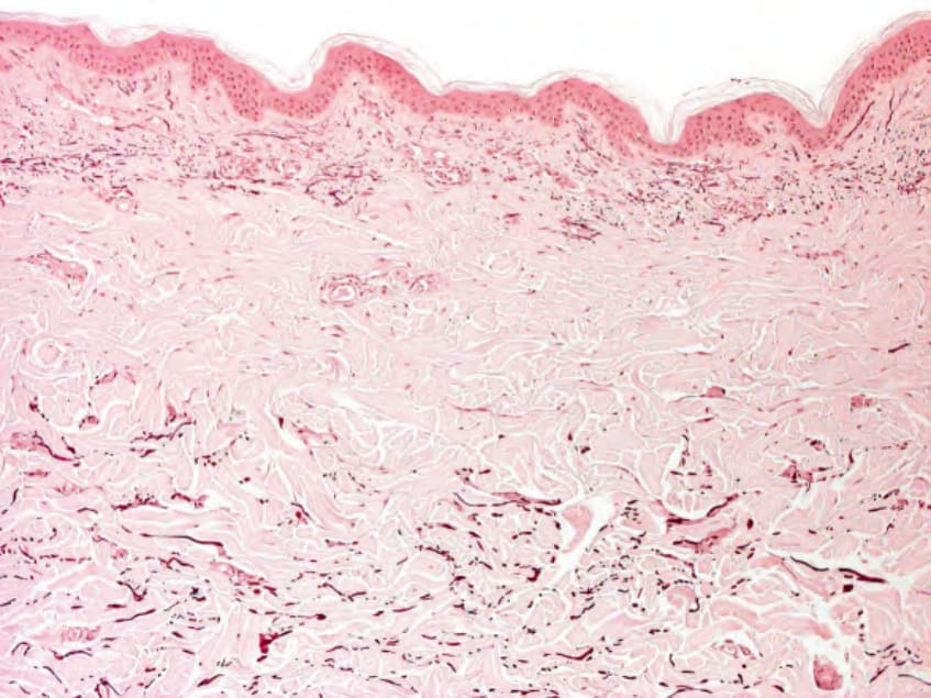
Fig. 99.2 Mid-dermal elastolysis – histopathologic features. A near absence of elastic fibers in a band-like distribution in the mid reticular dermis. Identification of these changes requires an elastic tissue stain (e.g. orcein). Courtesy Lorenzo Cerroni, MD.
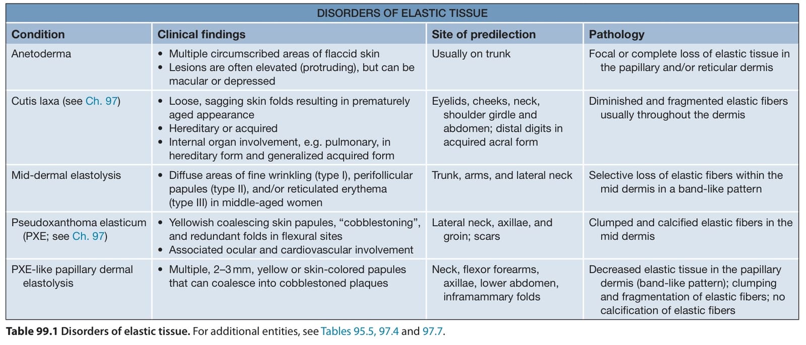
Table 99.1 Disorders of elastic tissue. For additional entities, see Tables 95.5, 97.4 and 97.7.