ANETODERMA
Synonyms: Macular atrophy Anetoderma maculosa Anetoderma maculosa cutis Atrophia maculosa cutis
Key features
Circumscribed 1–2 cm areas of flaccid skin, which may be elevated, macular, or depressed
Primary anetoderma has classically been divided into inflammatory and non-inflammatory
Secondary anetoderma is associated with infectious and inflammatory cutaneous disorders as well as tumors and autoimmune disorders (e.g. antiphospholipid antibody syndrome)
Histologically, focal absence of dermal elastic tissue is seen
Introduction
The term “anetoderma” is derived from anetos, the Greek word for slack, and derma for skin. Anetoderma is an elastolytic disorder characterized by localized areas of flaccid skin, which may be depressed, macular or papular; the latter can reflect herniation of the subcutaneous tissue. Anetoderma may be idiopathic or associated with an inflammatory disorder of the skin.
History
The first case of primary inflammatory anetoderma was reported by Jadassohn in 1892. A year previously, Schweninger and Buzzi described a 29-year-old woman with multiple, non-inflammatory, sac-like tumors on the trunk and upper extremities.
Epidemiology
Several hundred cases of anetoderma have been reported in the world literature. Primary anetoderma favors young adults between 15 and 25 years of age and occurs more frequently in women than men. The epidemiology of secondary anetoderma reflects that of the underlying etiologies (see Table 99.3).
Pathogenesis
The pathogenesis of anetoderma is not known. These lesions could be considered unusual scars, since scars also have decreased elastic tissue. The loss of dermal elastin may reflect an impaired turnover of elastin, caused by either increased destruction or decreased synthesis of elastic fibers. There are a number of proposed explanations for the focal elastin destruction, e.g. the release of elastase from inflammatory cells, the release of cytokines such as interleukin-6, an increased production of progelatinases A and B, and the phagocytosis of elastic fibers by macrophages. More recently, a decrease in fibulin protein expression was observed, suggesting defective elastic fiber reassembly. Immunologic mechanisms may also play a role, providing an explanation for associated findings such as antiphospholipid antibodies (see below). Based upon a few isolated patients, a link may exist between anetoderma and mutations in genes that encode enzymes in the citric acid cycle.
In primary anetoderma, there is no underlying associated disorder and lesions arise within clinically normal skin. It has classically been subdivided into two major forms: those with preceding inflammatory lesions (the Jadassohn–Pellizzari type) and those without preceding inflammatory lesions (the Schweninger–Buzzi type) (Table 99.2). This clinical classification is primarily of historical interest, since the two types of lesions can coexist in the same patient and their histopathology
is often the same; the presence or absence of clinical inflammation at the onset of the disease is not related to prognosis.
Secondary anetoderma can arise in the setting of a primary inflammatory dermatosis, skin infection or cutaneous tumor, as well as in patients with systemic disorders (e.g. autoimmune). Over the past decade, an association with antiphospholipid antibody syndrome has been highlighted. Although the vast majority of cases are sporadic, familial anetoderma has been described and is usually not associated with pre-existing lesions.
Clinical Features
The characteristic lesions are flaccid, circumscribed areas of slack skin that are a reflection of a marked reduction or absence of dermal elastic fibers; they can appear as depressions, wrinkling, or sac-like protrusions (Fig. 99.3). These atrophic lesions vary in number from a few to hundreds, and they typically measure 1–2 cm in diameter and are skin-colored to bluish–white in color. The skin surface can be normal in appearance or wrinkled, and a central depression may be seen. Coalescence of smaller lesions can give rise to larger herniations.
The examining finger sinks into a distinct pit with sharp borders as if into a hernia ring. The bulge reappears when the pressure from the finger is released. This clinical finding is referred to as the “buttonhole” sign and is similar to that observed in neurofibromas.
Predilection sites for these asymptomatic lesions are the chest, back, neck, and upper extremities. Primary anetoderma usually develops in young adults, and new lesions often continue to form for many years as the older lesions fail to resolve.
True secondary anetoderma implies that the characteristic atrophic lesion has appeared in the same site as a previous specific skin lesion, e.g. from varicella or leprosy. Some authors also consider lesions associated
with an underlying disease (e.g. HIV infection, antiphospholipid antibody syndrome, autoimmune thyroiditis) as secondary anetoderma; however, in this instance, the atrophic areas do not necessarily develop within areas of known inflammation. Evaluation for the presence of antiphospholipid antibodies should be performed before patients with idiopathic anetoderma are labeled as having primary anetoderma. The range of heterogeneous conditions associated with secondary anetoderma are outlined in Table 99.3. With the exception of distribution (and perhaps size), the clinical features are the same as those of primary anetoderma.
Anetoderma has increasingly been reported in premature infants. In most neonates, it appears to be related to the use of cutaneous monitoring leads or adhesives, as well as extreme prematurity.
In patients with anetoderma, a variety of systemic abnormalities have been reported, including ocular, endocrinologic, skeletal, cardiac, pulmonary, and gastrointestinal disease. Because there has been no consistency with regard to these associated abnormalities, they are probably coincidental, although the possibility exists that they may reflect a more generalized elastolytic disorder that has yet to be defined.
Pathology
In routinely stained sections, the collagen within the dermis of affected skin appears normal. Perivascular lymphocytes, primarily T helper cells, are often present but do not correlate with clinical findings of inflammation.
The predominant abnormality, revealed by elastic tissue stains, is a focal, more or less complete loss of elastic tissue in the papillary and/or reticular dermis. There are usually some residual abnormal, irregular, and fragmented elastic fibers (Fig. 99.4). In addition to plasma cells and histiocytes with phagocyted elastic fibers, occasional granuloma formation can be seen.
Direct immunofluorescence sometimes shows linear or granular deposits of immunoglobulins and complement along the dermal– epidermal junction or around the dermal blood vessels in affected skin. However, these findings are not helpful diagnostically.
By electron microscopy, the elastic fibers are fragmented and irregular in shape and occasionally they are engulfed by macrophages.
Differential diagnosis
Anetoderma must be differentiated from other disorders of elastic tissue such as MDE (see Table 99.1) as well as atrophodermas (see below). However, the major differential diagnosis consists of post-traumatic scars and papular elastorrhexis. The latter is an acquired disorder characterized by firm, white, non-follicular papules that measure 2–5 mm in diameter and are evenly scattered on the trunk. The lesions usually appear during adolescence or early adulthood. Histology demonstrates focal degeneration of elastic fibers and normal collagen. There are no associated extracutaneous abnormalities. This disorder is believed by some authors to be a variant of connective tissue nevi or an abortive form of the Buschke–Ollendorff syndrome, while others think
that these lesions represent papular acne scars. In contrast to anetoderma, lesions of papular elastorrhexis are firm and non-compressible.
Less often, anetoderma is confused with perifollicular elastolysis (see above), nevus lipomatosus, or focal dermal hypoplasia (Goltz
syndrome). Nevus lipomatosus superficialis of Hoffman and Zurhelle presents as a clustered group of soft, skin-colored to yellow nodules, usually located on the lower trunk and present since birth (see Ch. 117). Histology shows ectopic mature lipocytes located within the dermis. The lesions of Goltz syndrome (including telangiectasias, vermiculate dermal atrophy, hypopigmentation, hyperpigmentation, and fatty herniations/hamartomas) are in a linear array along the lines of Blaschko (see Ch. 62). Histologically, a decrease in dermal content is seen as well as extension or deposition of subcutaneous fat into the dermis.
Treatment
Various therapeutic modalities have been tried, but have not resulted in improvement of existing atrophic lesions; these include intra lesional triamcinolone and systemic administration of aspirin, dapsone, phenytoin, penicillin G, vitamin E, and inositol niacinate. Some authors have reported improvement with hydroxychloroquine. Surgical excision of limited lesions may be helpful, with informed consent from the patient regarding the risk of scar formation. The utility of soft tissue fillers is inconclusive. Based upon case reports, fractionated ablative and non-ablative lasers may be beneficial.
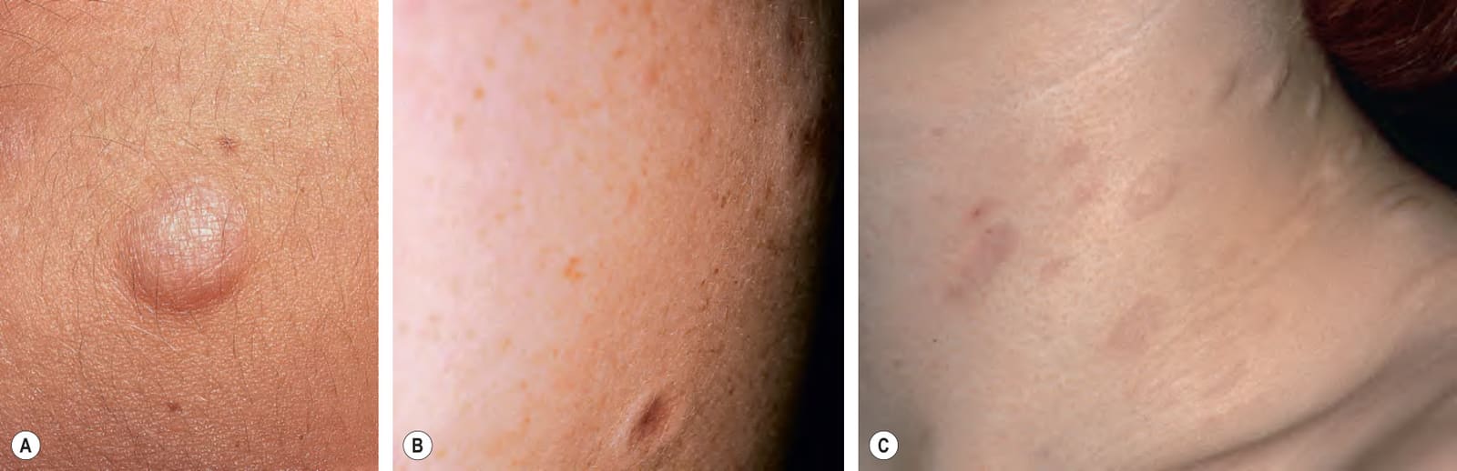
Fig. 99.3 Primary anetoderma. Lesions can range from soft, skin-colored papules that herniate upon palpation (A) to flaccid papules that have a central depression (B). The upper trunk and neck is a common location for primary anetoderma (C). A, Courtesy Ronald P. Rapini, MD; C, Courtesy Thomas Schwarz, MD.
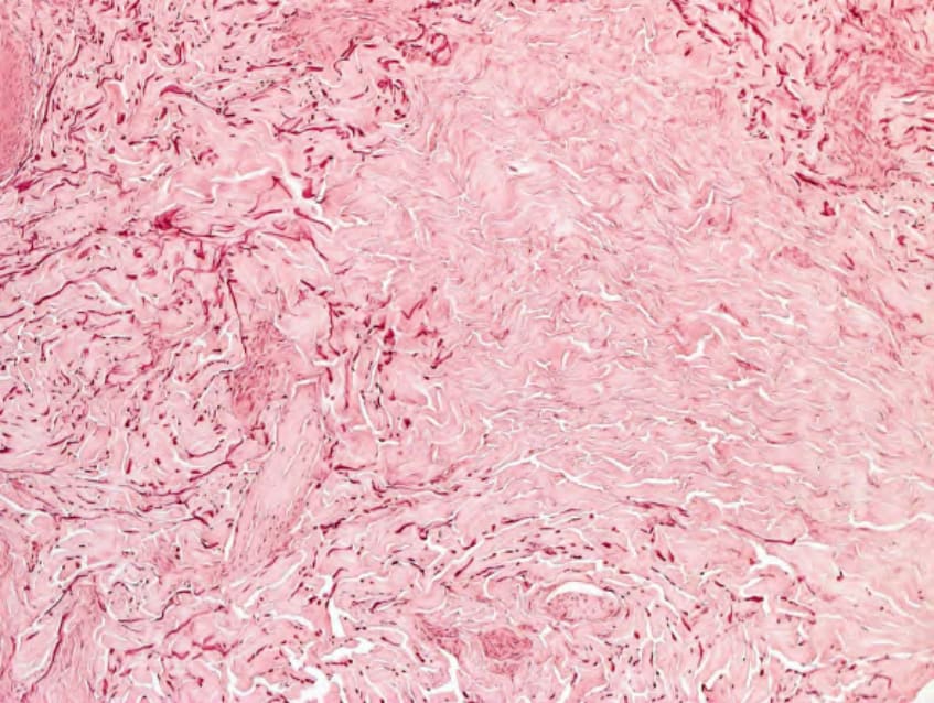
Fig. 99.4 Anetoderma – histopathologic features. A decrease of elastic fibers in both the papillary and reticular dermis (Weigert’s stain). Courtesy Lorenzo Cerroni, MD.
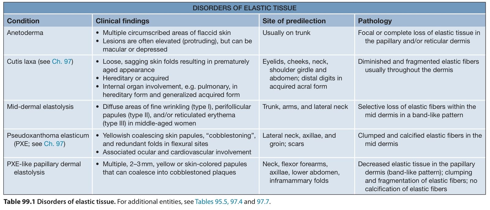
Table 99.1 Disorders of elastic tissue. For additional entities, see Tables 95.5, 97.4 and 97.7.
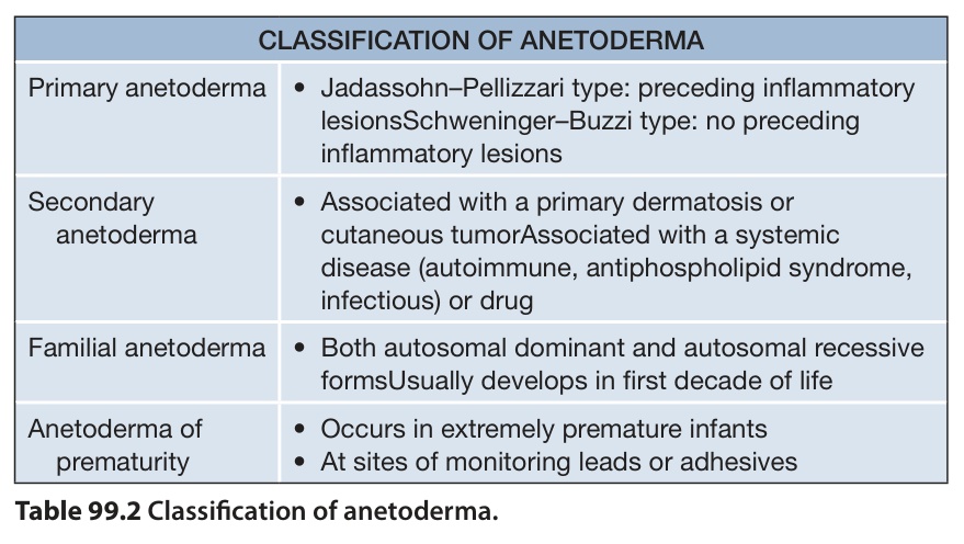
Table 99.2 Classification of anetoderma.
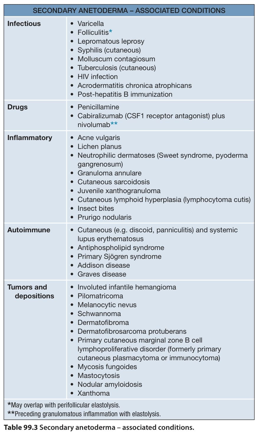
Table 99.3 Secondary anetoderma – associated conditions.