STRIAE
Synonyms: Striae distensae Striae atrophicans “Stretch marks” Striae gravidarum
Introduction
Striae are a very common condition in most age groups. They are linear atrophic depressions of the skin that form in areas of dermal damage produced by stretching of the skin. They are associated with various physiologic states, including puberty, pregnancy, rapid growth, weight gain or loss, obesity, and disorders that lead to hypercortisolism. Striae also develop at sites of potent topical corticosteroid application, in particular, occluded intertriginous zones.
History
Striae were first described by Roederer in 1773, and the first histologic descriptions were made by Troisier and Menetrier in 1889.
Epidemiology
Striae are very common and usually develop between the ages of 5 and 50 years. They are seen more commonly in White individuals and occur about twice as frequently in women as in men. They commonly develop during puberty, with a prevalence ranging from 5% to 85%, or during pregnancy, with an incidence of up to 90%.
Pathogenesis
Factors leading to the development of striae have not been fully elucidated. They are a reflection of “breaks” in the connective tissue that lead to dermal atrophy. A number of factors, including hormones (particularly corticosteroids), mechanical stress and genetic predisposition, appear to play a role. Bevacizumab, an anti-VEGF antibody well known for its negative effect on wound healing, has also been associated with severe ulcerations of corticosteroid-induced striae.
Clinical Features
Striae are usually multiple, symmetric, well-defined, linear atrophic lesions that often follow the lines of cleavage. They are usually more of a cosmetic concern, but rarely can ulcerate. Initially, striae appear as red-to-violaceous elevated lines that can be mildly pruritic and are called striae rubrae (Fig. 99.5). Over time, the color gradually fades, and the lesions become atrophic, exhibiting a fine wrinkled appearance. These striae albae are usually permanent, but they may fade somewhat over time. The striae can measure several centimeters in length and a few millimeters to a few centimeters in width.
During puberty, striae appear in areas where there is a rapid increase in size. In girls, the most common sites are the thighs, hips, buttocks and breasts, whereas in boys, they are seen on the shoulders, thighs, buttocks, and lumbosacral region (Fig. 99.6). Other less common sites include the abdomen, upper arms, neck, and axillae.
Striae distensae are a common finding on the abdomen, and less so on the breasts and thighs, of pregnant women, especially during the last trimester. They are more common in younger primigravidas than in older pregnant women, in those who gained more weight during pregnancy, and/or those who had babies with a higher birth weight. The development of striae gravidarum has been associated with an increased risk of laceration during vaginal delivery as well as subsequent pelvic relaxation and clinical prolapse.
The striae associated with systemic corticosteroid therapy and Cushing syndrome can be larger and more widely distributed (see Ch. 53). Flexural and intertriginous areas are particularly at risk for developing striae from the use of potent topical corticosteroids.
Atrophic striae may become elevated and “worm-like” in the setting of severe edema, including lymphedema. In polymorphic eruption of pregnancy, initial lesions often arise within striae gravidarum.
Pathology
Histologic findings are similar to those of scars and depend upon the stage of evolution of the striae at the time the biopsy is performed. The epidermis can be normal during the early stages, but eventually becomes atrophic with blunted rete ridges. The dermal thickness is decreased, as is the collagen in the upper dermis. The collagen bundles lie parallel to the epidermis. Alterations in elastic fibers are variable, but they can be fragmented, and specific elastin staining can demonstrate a marked reduction compared with adjacent normal dermis. There is an absence of both hair follicles and other adnexal structures.
Differential Diagnosis
The diagnosis of striae distensae is usually straightforward, but the differential diagnosis does include linear focal elastosis (elastotic striae), an entity first described by Burket et al. in 1989. Linear focal elastosis is characterized by rows of yellow, palpable, striae-like bands on the lower back. Unlike striae, the lesions are raised and yellow rather than depressed and white. Elderly men are most commonly affected. Histologically, there is a focal increase in the number of elongated or fragmented elastic fibers as well as a thickened dermis. It is postulated that linear focal elastosis may represent an excessive regenerative process of elastic fibers and could be viewed as a keloidal repair of striae distensae.
Treatment
Striae distensae have no medical consequences, but they are frequently distressing to those afflicted. As striae tend to improve spontaneously over time, the value of anecdotal therapies without case controls is difficult to assess. Topical treatments that have shown some benefit in early-stage striae include tretinoin 0.1% cream and a combination of 20% glycolic acid with either 0.05% tretinoin or 10% L-ascorbic acid.
The evidence for the effect of other topical treatments, either in prevention or treatment of striae, is inconclusive.
Several lasers have been used to treat striae: the 585 nm pulsed dye laser may improve the appearance of striae rubrae, but it has no effect on striae albae; secondary pigmentary alterations in darker skin are a potential complication. Improvement of striae albae was noted with fractionated non-ablative lasers, presumably via neocollagenesis. Use of radiofrequency and pulsed magnetic fields may also lead to improvement. However, currently, there is no specific therapy that results in a complete response.
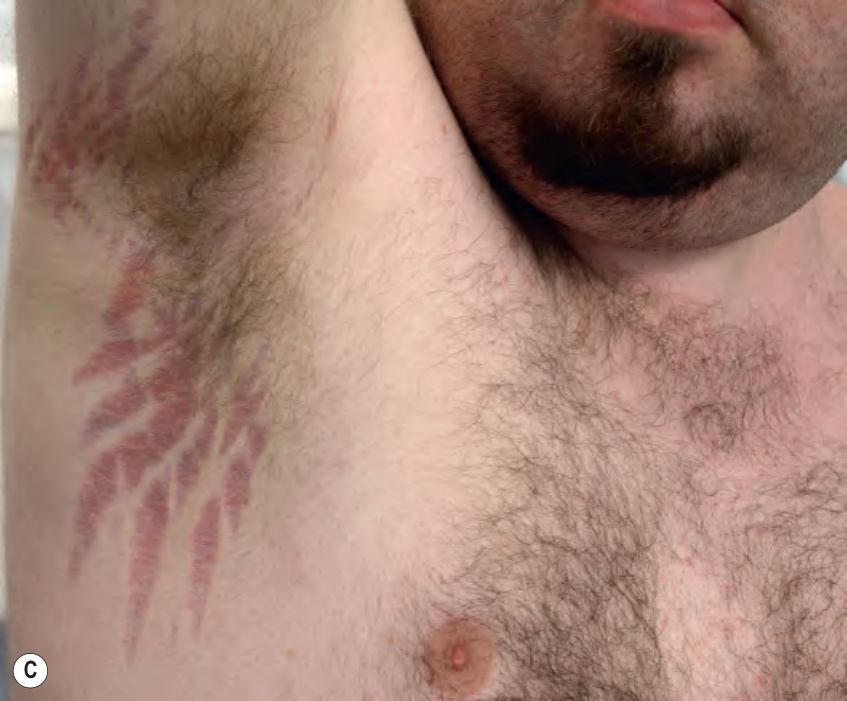
Fig. 99.5 Striae.A Linear erythematous lesions on the abdomen (striae rubrae). B Atrophic linear lesions of striae albae in a teenager. C Large axillary striae in a patient receiving chronic, high-dose systemic corticosteroids. B, Courtesy Kalman Watsky, MD.
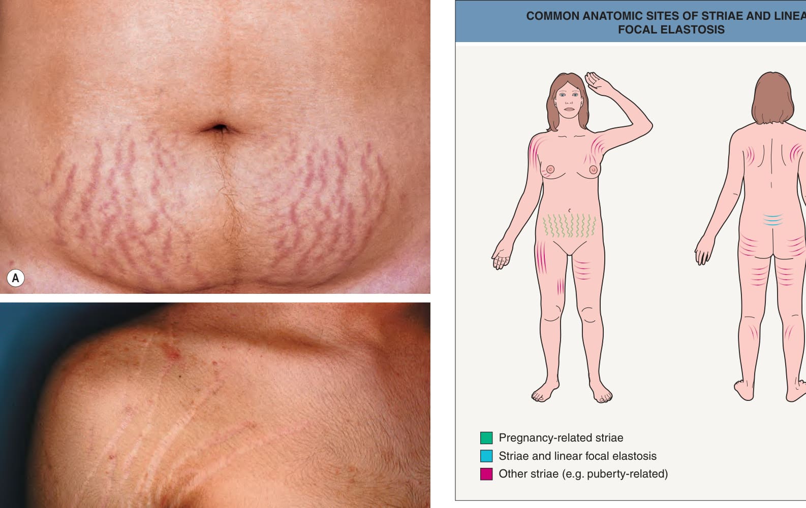
Fig. 99.6 Common anatomic sites of striae and linear focal elastosis. Striae associated with pregnancy are in green and lesions of linear focal elastosis are in blue.