FOLLICULAR ATROPHODERMA
Follicular atrophoderma refers to dimple-like depressions at the follicular orifices. It can occur as an isolated defect of limited extent, in association with a variety of disorders in which hair follicles are plugged with keratin, or with rare genodermatoses.
In 1944, Miescher described follicular atrophoderma in an 8-year-old girl with atypical chondrodystrophy. Six years later, Curth also used the term follicular atrophoderma (although there was no evidence of follicular atrophy but rather follicular agenesis) and further reported in 1978 on the genetics of follicular atrophoderma.
Clinical features
Distinctive follicular ice-pick depressions can be seen most commonly on the back of the hands and feet or on the cheeks. These pitted scars are often present at birth or appear during childhood. A family history may be present. Follicular atrophoderma can be associated with Bazex– Dupré–Christol and Conradi–Hünermann–Happle syndromes. When the lesions are found exclusively on the cheeks, the term “atrophoderma vermiculatum” applies. Atrophoderma vermiculatum can in turn be associated with various disorders discussed in the next section.
Atrophoderma Vermiculatum
Atrophoderma vermiculatum, a disorder limited to the face, has been described under a variety of names, including ulerythema acneiforme, acne vermoulante, atrophoderma reticulata symmetrica faciei, folliculitis ulerythema reticulata, folliculitis ulerythemosa, and honeycomb atrophy (“ulerythema” means scar plus redness and “vermiculatum” means worm-eaten).
Atrophoderma vermiculatum may: (1) occur sporadically; (2) be inherited as an autosomal dominant disorder; (3) be part of a group of related diseases that are referred to as “keratosis pilaris atrophicans” (see Table 38.2); or (4) be associated with various syndromes.
Multiple symmetric inflammatory papules on the cheeks, presumably centered around hair follicles, may precede the atrophic lesions. These papules then go on to become pitted, atrophic, depressed scars in a reticulated or honeycomb pattern (Fig. 99.8). The severity of erythema varies, as does the presence of milia and horny follicular plugs. Lesions can extend to the forehead and preauricular regions. Onset is usually in childhood or, less often, around puberty. Both sexes appear to be equally affected, and it usually has a slowly progressive course.
Atrophoderma vermiculatum can be associated with a group of closely related disorders referred to as keratosis pilaris atrophicans (see Table 38.2). This group also includes keratosis follicularis spinulosa decalvans and ulerythema ophryogenes. These conditions are characterized by keratotic follicular papules, variable degrees of inflammation, and secondary atrophic scarring. Ulerythema ophryogenes (or keratosis pilaris atrophicans faciei) differs from atrophoderma vermiculatum by affecting primarily the lateral portion of the eyebrows (ophryogenes) in the form of erythema, follicular papules, and alopecia. The inheritance pattern is autosomal dominant with incomplete penetrance and occasionally autosomal recessive, with progression usually ceasing after puberty. Keratosis follicularis spinulosa decalvans begins during
childhood as keratotic follicular papules on the malar area and progresses to involve the eyebrows, scalp, and extremities, with associated scarring alopecia. In most patients, this disorder is inherited in an X-linked recessive fashion.
The underlying pathogenesis in atrophoderma vermiculatum as well as the other disorders in the keratosis pilaris atrophicans group appears to be abnormal follicular hyperkeratinization. The latter occurs in the upper third of the hair follicle, leading to obstruction of the growing hair shaft and production of chronic inflammation. The end result is scarring below the level of obstruction.
Histopathology is usually not very helpful and shows dilated follicles, sometimes associated with plugging, inflammation, and sclerosis of dermal collagen.
Syndromes associated with atrophoderma vermiculatum include Rombo syndrome (milia, telangiectasias, basal cell carcinomas [BCCs], hypotrichosis, acrocyanosis, and, occasionally, trichoepitheliomas) and Loeys–Dietz syndrome (see Ch. 95), in addition to the less well characterized Nicolau–Balus syndrome (syringomas and milia), Tuzun syndrome, and Braun–Falco–Marghescu syndrome (congenital poikiloderma).
The differential diagnosis of atrophoderma vermiculatum includes atrophia maculosa varioliformis cutis, keratosis pilaris rubra of the cheeks, and, in older adults, erythromelanosis faciei.
This disorder is primarily a cosmetic problem. Various topical treatments, including emollients, corticosteroids, tretinoin and keratolytics, have shown no consistent benefit. In some instances, systemic isotretinoin has been shown to stop progression and to induce remission. Dermabrasion, laser therapy (e.g. CO, 585 nm pulsed dye), and fillers (e.g. hyaluronic acid, autologous fat) are other options for improving the appearance of the atrophic scars.
Bazex–Dupré–Christol Syndrome
Bazex, Dupré and Christol first described this genodermatosis in 1964, not to be confused with Bazex syndrome (acrokeratosis paraneoplastica) in which hyperkeratotic plaques of the ears, nose, cheeks, hands, feet and knees are associated with carcinomas of the upper aerodigestive tract (see Ch. 53). Bazex–Dupré–Christol syndrome is characterized by follicular atrophoderma, milia, multiple BCCs, hypotrichosis, and localized hypohidrosis (above the neck). It is inherited in an X-linked dominant fashion, and the gene has been linked to Xq24–q27. Additional reported findings include facial hyperpigmentation, hair shaft anomalies, and multiple trichoepitheliomas. Systemic manifestations are absent. Recently, it was suggested that this syndrome might better be classified as an ectodermal dysplasia.
The follicular atrophoderma, described as multiple ice-pick marks or patulous follicles, is found most commonly on the dorsal aspect of the hands, but can also be seen on the feet, lower back, elbows, and occasionally the face; it may be present at birth or appear later during childhood. No abnormalities of the elastic fibers have been found (nor any evidence of atrophy of the epidermis, hair or dermis), making the term “follicular atrophoderma” a misnomer. The histopathology usually shows hair follicles that are abnormally wide, plugged, and surrounded by an inflammatory cell infiltrate and clusters of basaloid cells. Sweat glands can be absent.
BCCs develop in about 40% of patients (primarily on the face) and can resemble melanocytic nevi; the age of onset varies from 9 to 50 years of age. To date, ~20 families with this syndrome have been described.
The differential diagnosis includes other genodermatoses with multi-ple BCCs: basal cell nevus syndrome (Gorlin syndrome), an autosomal dominant disorder most commonly due to mutations in PTCH1 (see Ch. 108); Rombo syndrome, an autosomal dominant disorder characterized by BCCs, atrophoderma vermiculatum, milia, hypotrichosis, acrocyanosis, and, occasionally, trichoepitheliomas; and xeroderma pigmentosum (see Ch. 86).
Conradi–Hünermann–Happle Syndrome (X-Linked Dominant Chondrodysplasia Punctata, CDPX2)
Conradi–Hünermann–Happle syndrome is an X-linked dominant disorder that occurs almost exclusively in girls, since it is usually lethal in hemizygous males. This form of chondrodysplasia punctata results from mosaicism for mutations in the gene on the X chromosome that encodes the emopamil-binding protein. The clinical manifestations include ichthyosiform streaks (composed of erythema plus feathery, adherent scale) patterned along the lines of Blaschko; they usually resolve during the first year of life and are replaced by bands of follicular atrophoderma. Hyperpigmentation, cataracts, scarring alopecia, saddle-nose deformity, asymmetric limb reduction defects, and stippled calcifications of the epiphyses are additional manifestations (see Ch. 57). Within the ichthyosiform areas in newborns, keratotic follicular plugs containing dystrophic calcification is a distinctive histopathologic feature.
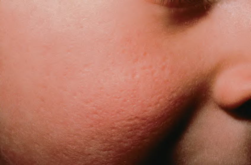
Fig. 99.8 Atrophoderma vermiculatum. Multiple small pitted scars on the cheek of a young girl. Note the honeycomb pattern on the lower inner cheek; the skin is said to appear “worm-eaten”. Courtesy Robert Hartman, MD.