PIEZOGENIC PEDAL PAPULES (PIEZOGENIC PAPULES)
Piezogenic pedal papules were first described by Shelley and Rawnsley in 1968. This term is somewhat of a misnomer as piezogenic means “producing pressure”, when in fact these lesions are produced by pressure. It is postulated that pressure induces herniation of fat through connective tissue in the dermis of the heels.
These papules are common enough in the general population to be considered normal and have also been described in association with Ehlers–Danlos syndrome, Prader–Willi syndrome, and GATA2 deficiency. An infantile variant characterized by larger nodules on the medial aspect of the heel (present without weight-bearing) has been described and is referred to as pedal papules of infancy and more recently, precalcaneal congenital fibrolipomatous hamartoma (Fig. 99.10). When unilateral, this entity has to be distinguished from juvenile aponeurotic fibroma which can calcify.
Skin-colored papules and nodules are seen on the sides of the heels (Fig. 99.11); they are induced by weight-bearing and disappear when the leg is
raised. The lesions are usually asymptomatic, but pain has been described during weight-bearing, and, in these cases, an orthopedic shoe or surgical excision may help. Similar lesions have been described on the wrists.
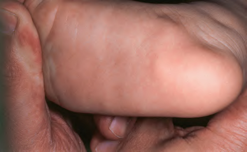
Fig. 99.10 Pedal papules of infancy (precalcaneal congenital fibrolipomatous hamartoma). Soft nodules on the medial and plantar surfaces of the foot. Courtesy Julie V. Schaffer, MD.
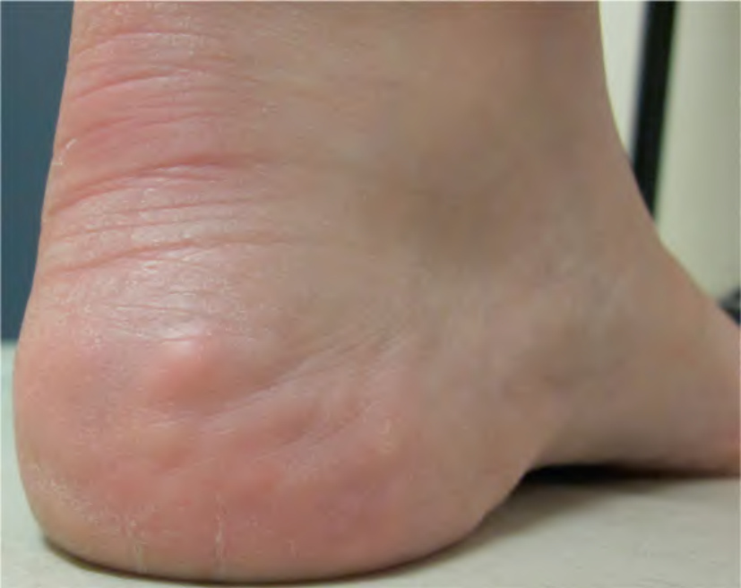
Fig. 99.11 Piezogenic pedal papules. Soft papulonodules on the heel. Courtesy Benoit Côté, MD.